Application
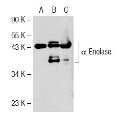
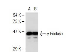
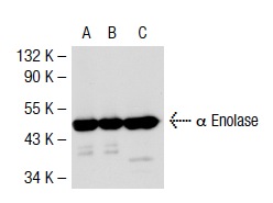
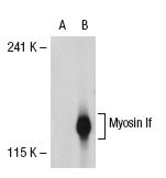
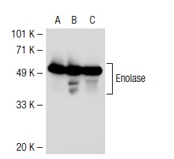
Western blot analysis of α Enolase expression in non-transfected 293T: (A), human α Enolase transfected 293T: (B) and Hep G2 (C) whole cell lysates.Application
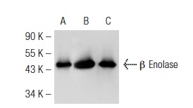
Western blot analysis of β Enolase expression in non-transfected 293T: (A), mouse β Enolase transfected 293T: (B) and Hep G2 (C) whole cell lysates.Application
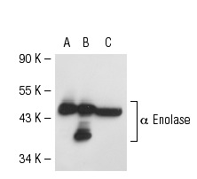
Western blot analysis of α Enolase expression in non-transfected 293T: (A), mouse α Enolase transfected 293T: (B) and NIH/3T3 (C) whole cell lysates.Application
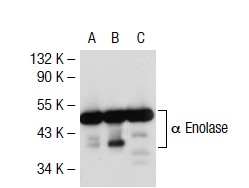
Western blot analysis of α Enolase expression in non-transfected 293T: (A), mouse α Enolase transfected 293T: (B) and A549 (C) whole cell lysates.Application
Western blot analysis of Enolase expression in non-transfected: (A) and mouse Enolase transfected: (B) 293T whole cell lysates.Application
Western blot analysis of α Enolase expression in non-transfected 293T: (A), mouse α Enolase transfected 293T: (B) and A549 (C) whole cell lysates.Application
Immunoperoxidase staining of formalin fixed, paraffin-embedded human bronchus tissue showing cytoplasmic staining of respiratory epithelial cells (low and high magnification). Kindly provided by The Swedish Human Protein Atlas (HPA) program.Application
Western blot analysis of Enolase expression in non-transfected 293T: (A), human Enolase transfected 293T: (B) and Hep G2 (C) whole cell lysates.Application
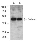
Western blot analysis of Enolase expression in A549 whole cell lysate (A) and rat skeletal muscle tissue extract (B).Application
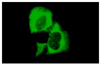
Immunofluorescence staining of methanol-fixed A549 cells showing cytoplasmic localization.| Product Name | Enolase Antibody (H-300) |
|---|---|
| Antibody Type | Primary Antibodies |
| Antigen Alias | raised against amino acids 1-300 mapping at the N-terminus of α Enolase of human origin |
| Modification Notes | Enolases have been characterized as highly conserved cytoplasmic glycolytic enzymes that may be involved in differentiation. Three isoenzymes have been identified: å enolase, ∫ enolase and © enolase. å enolase expression has been detected on most tissues, whereas ∫ enolase is expressed predominantly in muscle tissue and © enolase is detected only in nervous tissue. These isoforms exist as both homodimers and heterodimers, and they play a role in converting phosphoglyceric acid to phosphenolpyruvic acid in the glycolytic pathway. The 433 amino acid protein shows 67% homology to yeast enolase and 94% homology to rat nonneural enolase. Studies also indicate that å enolase is encoded by the same gene that encodes tau-crystallin, a lens structural protein. |
| Clonality | Polyclonal |
|---|---|
| Isotype | IgG |
| Host Species | Rabbit |
| Tested Applications | WBIPIFIHCELISA |
| recommended for detection of α Enolase, β Enolase and γ Enolase of mouse, rat and human origin by WB, IP, IF, IHC(P), and ELISA also reactive with additional species, including equine, bovine and avian: |
|
| Species Reactivity | HumanMouseRat |
| Concentration | 1mg/ml |
| Purification | Affinity purified |
| Alternative Names | raised against amino acids 1-300 mapping at the N-terminus of &alpha Enolase of human origin |
|---|---|
| Tissue Specificity | raised against amino acids 1-300 mapping at the N-terminus of α Enolase of human origin |
| Entrez Gene | 2023 |
|---|
Application
Western blot analysis of α Enolase expression in non-transfected 293T: (A), human α Enolase transfected 293T: (B) and Hep G2 (C) whole cell lysates.
Application
Western blot analysis of β Enolase expression in non-transfected 293T: (A), mouse β Enolase transfected 293T: (B) and Hep G2 (C) whole cell lysates.
Application
Western blot analysis of α Enolase expression in non-transfected 293T: (A), mouse α Enolase transfected 293T: (B) and NIH/3T3 (C) whole cell lysates.
Application
Western blot analysis of α Enolase expression in non-transfected 293T: (A), mouse α Enolase transfected 293T: (B) and A549 (C) whole cell lysates.
Application
Western blot analysis of Enolase expression in non-transfected: (A) and mouse Enolase transfected: (B) 293T whole cell lysates.
Application
Western blot analysis of α Enolase expression in non-transfected 293T: (A), mouse α Enolase transfected 293T: (B) and A549 (C) whole cell lysates.
Application
Immunoperoxidase staining of formalin fixed, paraffin-embedded human bronchus tissue showing cytoplasmic staining of respiratory epithelial cells (low and high magnification). Kindly provided by The Swedish Human Protein Atlas (HPA) program.
Application
Western blot analysis of Enolase expression in non-transfected 293T: (A), human Enolase transfected 293T: (B) and Hep G2 (C) whole cell lysates.
Application
Western blot analysis of Enolase expression in A549 whole cell lysate (A) and rat skeletal muscle tissue extract (B).
Application
Immunofluorescence staining of methanol-fixed A549 cells showing cytoplasmic localization.| Application Notes | recommended for detection of α Enolase, β Enolase and γ Enolase of mouse, rat and human origin by WB, IP, IF, IHC(P), and ELISA also reactive with additional species, including equine, bovine and avian: |
|---|
| Form | Liquid |
|---|---|
| Storage Instructions | For short-term storage, store at 4° C. For long-term storage, aliquot and store at -20ºC or below. Avoid multiple freeze-thaw cycles. |
| Storage Buffer | phosphate buffered saline , pH 7.4, 150mM NaCl, 0.02% sodium azide and 50% glycerol. |


