Application
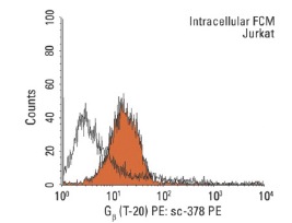
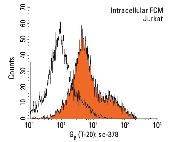
Indirect, intracellular FCM analysis of fixed and permeabilized Jurkat cells stained with Gb (T-20), followed by PE-conjugated mouse anti-rabbit IgG: . Black line histogram represents the isotype control, normal rabbit IgG: .Application
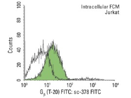
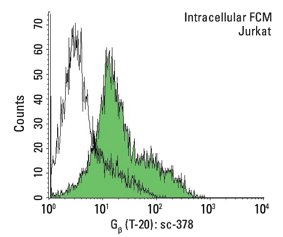
Indirect, intracellular FCM analysis of fixed and permeabilized Jurkat cells stained with Gb (T-20), followed by FITC-conjugated donkey anti-rabbit IgG: . Black line histogram represents the isotype control, normal rabbit IgG: .Application
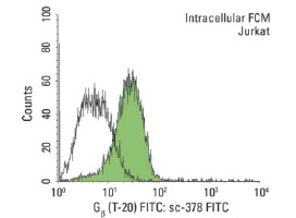
Indirect, intracellular FCM analysis of fixed and permeabilized Jurkat cells stained with Gb (T-20), followed by FITC-conjugated chicken anti-rabbit IgG: . Black line histogram represents the isotype control, normal rabbit IgG: .Application
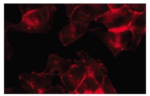
Immunofluorescence staining of methanol-fixed HeLa cells showing membrane localization.Application
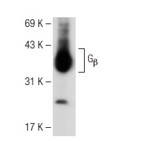
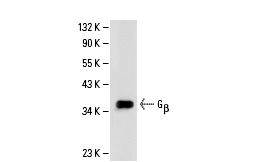
Western blot analysis of Gβ expression in Y79 whole cell lysate.Application
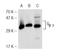
Western blot analysis of Gβ 3 expression in non-transfected 293T: (A), mouse Gβ 3 transfected 293T: (B) and Jurkat (C) whole cell lysates.Application
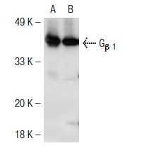
Western blot analysis of Gβ 1 expression in non-transfected: (A) and mouse Gβ 1 transfected: (B) 293T whole cell lysates.Application
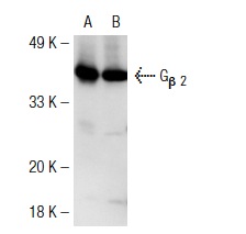
Western blot analysis of Gβ 2 expression in non-transfected: (A) and mouse Gβ 2 transfected: (B) 293T whole cell lysates.Application
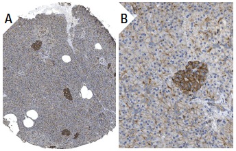
Immunoperoxidase staining of formalin fixed, paraffin-embedded human pancreas tissue showing membrane staining of Islet cells and exocrine glandular cells at low (A) and high (B) magnification. Kindly provided by The Swedish Human Protein Atlas (HPA) program.Application
Western blot analysis of Gβ expression in HeLa whole cell lysate.Application
Indirect, intracellular FCM analysis of fixed and permeabilized Jurkat cells stained with Gβ (T-20), followed by PE-conjugated goat anti-rabbit IgG: . Black line histogram represents the isotype control, normal rabbit IgG: .Application
Indirect, intracellular FCM analysis of fixed and permeabilized Jurkat cells stained with Gβ (T-20), followed by FITC-conjugated goat anti-rabbit IgG: . Black line histogram represents the isotype control, normal rabbit IgG: .Application
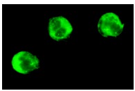
Immunofluorescence staining of methanol-fixed Jurkat cells showing cytoplasmic and membrane staining.Application
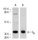
Western blot analysis of Gβ expression in Jurkat (A) whole cell lysate and bovine brain (B) extract.| Product Name | Gβ Antibody (T-20) |
|---|---|
| Antibody Type | Primary Antibodies |
| Antigen Alias | epitope mapping at the C-terminus of Gβ of mouse origin |
| Modification Notes | Heterotrimeric G proteins function to relay information from cell surface receptors to intracellular effectors (1). Each of a very broad range of receptors specifically detects an extracellular stimulus (a photon, pheromone, odorant, hormone or neurotransmitter) while the effectors (e.g., adenyl cyclase), which act to generate one or more intracellular messengers, are less numerous. In mammals, G protein å, ∫ and © polypeptides are encoded by at least 16, 4 and 7 genes, respectively (2-4). Most interest in G proteins has been focused on their å subunits, since these proteins bind and hydrolyze GTP and most obviously regulate the activity of the best studied effectors. Evidence, however, has established an important regulatory role for the ∫© subunits (5-7). It is becoming increasingly clear that different G protein complexes expressed in different tissues carry structurally distinct members of the © as well as the å and ∫ subunits and that preferential associations between members of subunit families increase G protein functional diversity (8,9). |
| Clonality | Polyclonal |
|---|---|
| Isotype | IgG |
| Host Species | Rabbit |
| Tested Applications | WBIPIFIHCFCELISA |
| recommended for detection of Gβ 1, Gβ2, Gβ 3 and Gβ 4 of mouse, rat, human and bovine origin by WB, IP, IF, IHC(P), ELISA and indirect FCM using PE (sc-3739)- and FITC (sc-2012)-conjugated goat anti-rabbit IgG also reactive with additional species, including equine, canine, bovine, porcine and avian: |
|
| Species Reactivity | HumanMouseRat |
| Concentration | 1mg/ml |
| Purification | Affinity purified |
| Alternative Names | epitope mapping at the C-terminus of G&beta of mouse origin |
|---|---|
| Tissue Specificity | epitope mapping at the C-terminus of Gβ of mouse origin |
| Entrez Gene | 59345 |
|---|
Application
Indirect, intracellular FCM analysis of fixed and permeabilized Jurkat cells stained with Gb (T-20), followed by PE-conjugated mouse anti-rabbit IgG: . Black line histogram represents the isotype control, normal rabbit IgG: .
Application
Indirect, intracellular FCM analysis of fixed and permeabilized Jurkat cells stained with Gb (T-20), followed by FITC-conjugated donkey anti-rabbit IgG: . Black line histogram represents the isotype control, normal rabbit IgG: .
Application
Indirect, intracellular FCM analysis of fixed and permeabilized Jurkat cells stained with Gb (T-20), followed by FITC-conjugated chicken anti-rabbit IgG: . Black line histogram represents the isotype control, normal rabbit IgG: .
Application
Immunofluorescence staining of methanol-fixed HeLa cells showing membrane localization.
Application
Western blot analysis of Gβ expression in Y79 whole cell lysate.
Application
Western blot analysis of Gβ 3 expression in non-transfected 293T: (A), mouse Gβ 3 transfected 293T: (B) and Jurkat (C) whole cell lysates.
Application
Western blot analysis of Gβ 1 expression in non-transfected: (A) and mouse Gβ 1 transfected: (B) 293T whole cell lysates.
Application
Western blot analysis of Gβ 2 expression in non-transfected: (A) and mouse Gβ 2 transfected: (B) 293T whole cell lysates.
Application
Immunoperoxidase staining of formalin fixed, paraffin-embedded human pancreas tissue showing membrane staining of Islet cells and exocrine glandular cells at low (A) and high (B) magnification. Kindly provided by The Swedish Human Protein Atlas (HPA) program.
Application
Western blot analysis of Gβ expression in HeLa whole cell lysate.
Application
Indirect, intracellular FCM analysis of fixed and permeabilized Jurkat cells stained with Gβ (T-20), followed by PE-conjugated goat anti-rabbit IgG: . Black line histogram represents the isotype control, normal rabbit IgG: .
Application
Indirect, intracellular FCM analysis of fixed and permeabilized Jurkat cells stained with Gβ (T-20), followed by FITC-conjugated goat anti-rabbit IgG: . Black line histogram represents the isotype control, normal rabbit IgG: .
Application
Immunofluorescence staining of methanol-fixed Jurkat cells showing cytoplasmic and membrane staining.
Application
Western blot analysis of Gβ expression in Jurkat (A) whole cell lysate and bovine brain (B) extract.| Application Notes | recommended for detection of Gβ 1, Gβ2, Gβ 3 and Gβ 4 of mouse, rat, human and bovine origin by WB, IP, IF, IHC(P), ELISA and indirect FCM using PE (sc-3739)- and FITC (sc-2012)-conjugated goat anti-rabbit IgG also reactive with additional species, including equine, canine, bovine, porcine and avian: |
|---|
| Form | Liquid |
|---|---|
| Storage Instructions | For short-term storage, store at 4° C. For long-term storage, aliquot and store at -20ºC or below. Avoid multiple freeze-thaw cycles. |
| Storage Buffer | phosphate buffered saline , pH 7.4, 150mM NaCl, 0.02% sodium azide and 50% glycerol. |


