Application
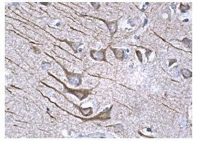
Immunoperoxidase staining of formalin fixed, paraffin-embedded human brain tissue showing cytoplasmic staining of neuronal cells.Application
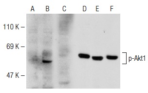
Antibodies tested include p-Akt1 (104A282): (A,B,C) and Akt1/2/3 (H-136): (D,E,F).Application
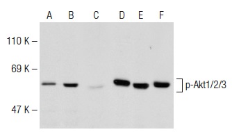
Antibodies tested include p-Akt1/2/3 (Ser 473): (A,B,C) and Akt1/2/3 (H-136): (D,E,F).Application
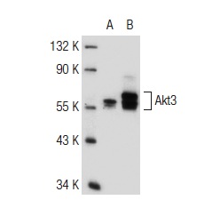
Western blot analysis of Akt3 expression in non-transfected: (A) and human Akt3 transfected: (B) 293 whole cell lysates.Application
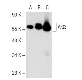
Western blot analysis of Akt3 expression in non-transfected 293: (A), human Akt3 transfected 293: (B) and IMR-32 (C) whole cell lysates.Application
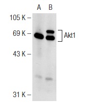
Western blot analysis of Akt1 expression in non-transfected: (A) and human Akt1 transfected: (B) 293T whole cell lysates.Application
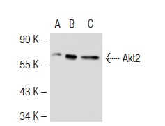
Western blot analysis of Akt2 expression in non-transfected 293T: (A), mouse Akt2 transfected 293T: (B) and KNRK (C) whole cell lysates.Application
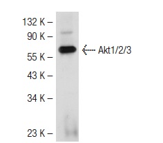
Western blot analysis of Akt1/2/3 expression in mouse brain tissue extract.Application
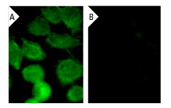
Immunofluorescence staining of methanol-fixed, control HeLa (A) and Akt1 siRNA silenced HeLa (B) cells showing diminished nuclear staining in the siRNA silenced cells. Cells probed with Akt1/2 (H-136): .Application
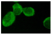
Immunofluorescence staining of methanol-fixed K-562 cells showing cytoplasmic and nuclear staining.Application
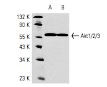
Western blot analysis of Akt1/2 expression in KNRK (A) and NIH/3T3 (B) whole cell lysates.| Product Name | Akt1/2/3 Antibody (H-136) |
|---|---|
| Antibody Type | Primary Antibodies |
| Antigen Alias | epitope corresponding to amino acids 345-480 of Akt1 of human origin |
| Conjugation | Agarose |
| Modification | Acetylated |
| Modification Notes | The serine/threonine kinase Akt family contains several members, including Akt1 (also designated PKB or RacPK), Akt2 (also designated PKB∫ or RacPK-∫) and Akt 3 (also designated PKB© or thyoma viral proto-oncogene 3), which exhibit sequence homology with the protein kinase A and C families and are encoded by the c-Akt proto-oncogene. All members of the Akt family have a pleckstrin homology domain. Akt1 and Akt2 are activated by PDGF stimulation. This activation is dependent on PDGFR-∫ tyrosine residues 740 and 751, which bind the subunit of the phosphatidylinositol 3-kinase (PI 3-kinase) complex. Activation of Akt1 by insulin or insulin-growth factor-1(IGF-1) results in phosphorylation of both Thr 308 and Ser 473. Akt proteins become phosphorylated and activated in insulin/IGF-1-stimulated cells by an upstream kinase(s), and the activation of Akt1 and Akt2 is inhibited by the PI kinase inhibitor wortmannin. Taken together, this data strongly suggests that the protein signals downstream of the PI kinases. Akt3 is phosphorylated on a serine residue in response to insulin. However, the activation of Akt3 by insulin is inhibited by prior activation of protein kinase C via a mechanism that does not require the presence of the PH domain. Akt3 is expressed in 3T3-L1 fibroblasts, adipocytes and skeletal muscle and may be involved in various biological processes, including adipocyte and muscle differentiation, glycogen synthesis, glucose uptake, apoptosis and cellular proliferation. |
| Clonality | Polyclonal |
|---|---|
| Isotype | IgG |
| Host Species | Rabbit |
| Tested Applications | ELISAIFIHCIPWB |
| WB:1:100~1:500 IP:1:20~1:100 IF:1:20~1:100 IHC-p:1:10~1:50 |
|
| Species Reactivity | HumanMouseRat |
| Predicted Reactivity | equine, canine, bovine, porcine and avian |
| Concentration | 1mg/ml |
| Purification | Affinity purified |
| Alternative Names | epitope corresponding to amino acids 345-480 of Akt1 of human origin |
|---|---|
| Tissue Specificity | epitope corresponding to amino acids 345-480 of Akt1 of human origin |
| Entrez Gene | 207 |
|---|
Application
Immunoperoxidase staining of formalin fixed, paraffin-embedded human brain tissue showing cytoplasmic staining of neuronal cells.
Application
Antibodies tested include p-Akt1 (104A282): (A,B,C) and Akt1/2/3 (H-136): (D,E,F).
Application
Antibodies tested include p-Akt1/2/3 (Ser 473): (A,B,C) and Akt1/2/3 (H-136): (D,E,F).
Application
Western blot analysis of Akt3 expression in non-transfected: (A) and human Akt3 transfected: (B) 293 whole cell lysates.
Application
Western blot analysis of Akt3 expression in non-transfected 293: (A), human Akt3 transfected 293: (B) and IMR-32 (C) whole cell lysates.
Application
Western blot analysis of Akt1 expression in non-transfected: (A) and human Akt1 transfected: (B) 293T whole cell lysates.
Application
Western blot analysis of Akt2 expression in non-transfected 293T: (A), mouse Akt2 transfected 293T: (B) and KNRK (C) whole cell lysates.
Application
Western blot analysis of Akt1/2/3 expression in mouse brain tissue extract.
Application
Immunofluorescence staining of methanol-fixed, control HeLa (A) and Akt1 siRNA silenced HeLa (B) cells showing diminished nuclear staining in the siRNA silenced cells. Cells probed with Akt1/2 (H-136): .
Application
Immunofluorescence staining of methanol-fixed K-562 cells showing cytoplasmic and nuclear staining.
Application
Western blot analysis of Akt1/2 expression in KNRK (A) and NIH/3T3 (B) whole cell lysates.| Application Notes | WB:1:100~1:500 IP:1:20~1:100 IF:1:20~1:100 IHC-p:1:10~1:50 |
|---|
| Form | Liquid |
|---|---|
| Storage Instructions | For short-term storage, store at 4° C. For long-term storage, aliquot and store at -20ºC or below. Avoid multiple freeze-thaw cycles. |
| Storage Buffer | phosphate buffered saline , pH 7.4, 150mM NaCl, 0.02% sodium azide and 50% glycerol. |


