Application
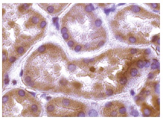
Immunoperoxidase staining of formalin fixed, paraffin-embedded human kidney tissue showing cytoplasmic staining of cells in tubules.Application
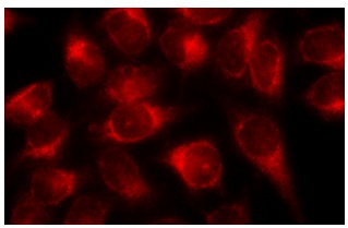
Immunofluorescence staining of methanol-fixed HeLa cells showing cytoplasmic localization.Application
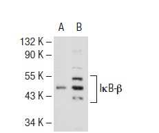
Western blot analysis of IκB-β expression in non-transfected: (A) and human IκB-β transfected: (B) 293T whole cell lysates.Application
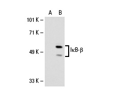
Western blot analysis of IκB-β expression in non-transfected: (A) and human IκB-β transfected: (B) 293 whole cell lysates.Application
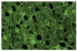
Immunofluorescence staining of normal mouse liver frozen section showing cytoplasmic staining.Application
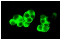
Immunofluorescence staining of methanol-fixed WEHI-231 cells showing cytoplasmic localization.Application
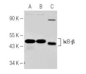
Western blot analysis of IκB-β expression in Raw 264.7 (A), WEHI-231 (B) and Ramos (C) whole cell lysates.Application
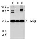
Western blot analysis of IκB-β expression in CTLL-2 (A), RAW 264.7 (B) and WEHI-231 (C) whole cell lysates.| Product Name | IκB-β Antibody (C-20) FITC |
|---|---|
| Antibody Type | Primary Antibodies |
| Antigen Alias | epitope mapping at the C-terminus of IκB-β of mouse origin |
| Modification Notes | On the basis of both functional and structural considerations, members of the I˚B family of proteins can be divided into four groups. The first of these groups, I˚B-å, includes the avian protein pp40 and the mammalian MAD-3, both of which inhibit binding of p50-p65 NFkB complex or Rel protein to their cognate binding sites but do not inhibit the binding of p50 homodimer to ˚B sites, suggesting that the I˚B-å family binds to the p65 subunit of p50-p65 heterocomplex through ankyrin repeats. The second member of the I˚B family is represented by a protein designated I˚B-∫. The third group of I˚B proteins is represented by I˚B-©, which is identical in sequence with the C-terminal domain of the p110 precursor of NFkB p50 and is expressed predominantly in lymphoid cells. An additional I˚B family member, I˚B-é, has several phosphorylated forms and is primarily found complexed with Rel A and/or c-Rel. |
| Clonality | Polyclonal |
|---|---|
| Isotype | IgG |
| Host Species | Rabbit |
| Tested Applications | WBIPIFIHCELISA |
| recommended for detection of IκB-β of mouse, rat and human origin by WB, IP, IF, IHC(P) and ELISA: |
|
| Species Reactivity | HumanMouseRat |
| Concentration | 1mg/ml |
| Purification | Affinity purified |
| Alternative Names | epitope mapping at the C-terminus of I&kappa B-&beta of mouse origin |
|---|---|
| Tissue Specificity | epitope mapping at the C-terminus of IκB-β of mouse origin |
| Entrez Gene | 4793 |
|---|
Application
Immunoperoxidase staining of formalin fixed, paraffin-embedded human kidney tissue showing cytoplasmic staining of cells in tubules.
Application
Immunofluorescence staining of methanol-fixed HeLa cells showing cytoplasmic localization.
Application
Western blot analysis of IκB-β expression in non-transfected: (A) and human IκB-β transfected: (B) 293T whole cell lysates.
Application
Western blot analysis of IκB-β expression in non-transfected: (A) and human IκB-β transfected: (B) 293 whole cell lysates.
Application
Immunofluorescence staining of normal mouse liver frozen section showing cytoplasmic staining.
Application
Immunofluorescence staining of methanol-fixed WEHI-231 cells showing cytoplasmic localization.
Application
Western blot analysis of IκB-β expression in Raw 264.7 (A), WEHI-231 (B) and Ramos (C) whole cell lysates.
Application
Western blot analysis of IκB-β expression in CTLL-2 (A), RAW 264.7 (B) and WEHI-231 (C) whole cell lysates.| Application Notes | recommended for detection of IκB-β of mouse, rat and human origin by WB, IP, IF, IHC(P) and ELISA: |
|---|
| Form | Liquid |
|---|---|
| Storage Instructions | For short-term storage, store at 4° C. For long-term storage, aliquot and store at -20ºC or below. Avoid multiple freeze-thaw cycles. |
| Storage Buffer | phosphate buffered saline , pH 7.4, 150mM NaCl, 0.02% sodium azide and 50% glycerol. |


