Application
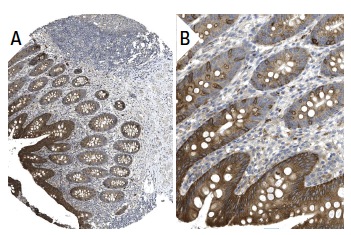
Immunoperoxidase staining of formalin fixed, paraffin-embedded human appendix tissue showing cytoplasmic and membrane staining of glandular cells at low (A) and high (B) magnification. Kindly provided by The Swedish Human Protein Atlas (HPA) program.Application
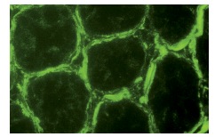
Immunofluorescence staining of normal mouse intestine frozen section showing membrane staining.Application
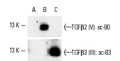
Western blotting analysis of 10 ng each of human recombinant TGFβ1 (A), TGFβ2 (B) and TGFβ3 (C).| Product Name | TGFβ3 (III) Antibody |
|---|---|
| Antibody Type | Primary Antibodies |
| Antigen Alias | epitope mapping near the C-terminus of TGFβ3 of human origin |
| Modification Notes | Transforming growth factor ∫s (TGF∫s) were originally discovered due to their ability to promote anchorage-independent growth of rat NRK fibroblasts in the presence of TGF∫. TGF∫1, TGF∫2 and TGF∫3 are each synthesized as precursor proteins that are very similar in that each is cleaved to yield a 112 amino acid polypeptide that remains associated with the latent portion of the molecules. TGF∫3 mediates many intercellular interactions that occur during embryonic development, cell differentiation and epithelial homeostasis. TGF∫3 overexpresses in extramammary Paget’s disease (EPD) and downregulates in Bowen’s disease, indicating that its expression is a useful indicator of tumor activity. TGF∫3 levels strongly correlate with IGF-1 and osteocalcin levels in serum. Significant amounts of TGF∫3 circulation appear to be representative of TGF∫3 expression in bone and may in part be derived from bone. Glucocorticoids may block TGF∫ production by modulating mRNA levels and c-Jun activity. |
| Clonality | Polyclonal |
|---|---|
| Isotype | IgG |
| Host Species | Rabbit |
| Tested Applications | WBIPIFIHCELISA |
| recommended for detection of TGFβ1, TGFβ2 and TGFβ3 of mouse, rat, human and avian origin by WB, IP, IF, IHC(P) and ELISA also reactive with additional species, including equine, canine, porcine and avian: |
|
| Species Reactivity | HumanMouseRat |
| Concentration | 1mg/ml |
| Purification | Affinity purified |
| Alternative Names | epitope mapping near the C-terminus of TGF&beta 3 of human origin |
|---|---|
| Tissue Specificity | epitope mapping near the C-terminus of TGFβ3 of human origin |
| Entrez Gene | 7043 |
|---|
Application
Immunoperoxidase staining of formalin fixed, paraffin-embedded human appendix tissue showing cytoplasmic and membrane staining of glandular cells at low (A) and high (B) magnification. Kindly provided by The Swedish Human Protein Atlas (HPA) program.
Application
Immunofluorescence staining of normal mouse intestine frozen section showing membrane staining.
Application
Western blotting analysis of 10 ng each of human recombinant TGFβ1 (A), TGFβ2 (B) and TGFβ3 (C).| Application Notes | recommended for detection of TGFβ1, TGFβ2 and TGFβ3 of mouse, rat, human and avian origin by WB, IP, IF, IHC(P) and ELISA also reactive with additional species, including equine, canine, porcine and avian: |
|---|
| Form | Liquid |
|---|---|
| Storage Instructions | For short-term storage, store at 4° C. For long-term storage, aliquot and store at -20ºC or below. Avoid multiple freeze-thaw cycles. |
| Storage Buffer | phosphate buffered saline , pH 7.4, 150mM NaCl, 0.02% sodium azide and 50% glycerol. |


