Application
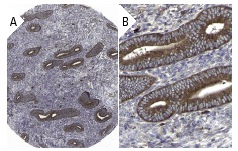
Immunoperoxidase staining of formalin fixed, paraffin-embedded human endometrium tissue showing cytoplasmic staining of glandular cells (low and high magni?fication). Kindly provided by The Swedish Human Protein Atlas (HPA) program.Application
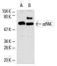
Western blot analysis of αPAK expression in mouse brain (A) and rat brain (B) extracts.Application
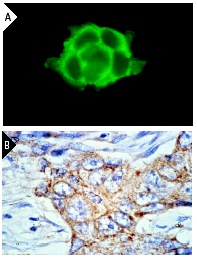
Immunofluorescence staining of methanol-fixed A-431 cells (A) and immunoperoxidase staining of formalin-fixed, paraffin-embedded human breast carcinoma tissue (B) showing cytoskeletal staining.| Product Name | αPAK Antibody (C-19) |
|---|---|
| Antibody Type | Primary Antibodies |
| Antigen Alias | epitope mapping at the C-terminus of αPAK of rat origin |
| Modification Notes | Three isoforms of serine/threonine kinases, designated åPAK p68, ∫PAK p65 and ©PAK p62, have been shown to exhibit a high degree of sequence homology with the S. cerevisiae kinase Ste 20, involved in pheromone signaling. The å, ∫ and ©PAK isoforms complex specifically with Rac1 and Cdc42 in their active GTP-bound state, inhibiting their intrinsic GTPase activity leading to their autophosphorylation. There are eight sites of autophosphorylation on ©PAK, including Ser 19, Ser 141 and Thr 402, and phosphorylation of Ser 141 and Thr 402 is correlated with ©PAK activation. Once phosphorylated and their affinity for Rac/Cdc42 reduced, the PAK isoforms disassociate from the complex to seek downstream substrates. One such putative substrate is Mek kinase, an upstream effector of Mek4 which is involved in the JNK signaling pathway. While the PAK isoforms interact in a GTP-dependent manner with Rac1 and Cdc42, they do not interact with Rho. |
| Clonality | Polyclonal |
|---|---|
| Isotype | IgG |
| Host Species | Rabbit |
| Tested Applications | WBIPIFIHCELISA |
| recommended for detection of αPAK p68 of mouse, rat and human origin by WB, IP, IF, IHC(P) and ELISA partially cross-reactive with γPAK and βPAK also reactive with additional species, including equine, canine, bovine, porcine and avian: |
|
| Species Reactivity | HumanMouseRat |
| Concentration | 1mg/ml |
| Purification | Affinity purified |
| Alternative Names | epitope mapping at the C-terminus of &alpha PAK of rat origin |
|---|---|
| Tissue Specificity | epitope mapping at the C-terminus of αPAK of rat origin |
| Entrez Gene | 5058 |
|---|
Application
Immunoperoxidase staining of formalin fixed, paraffin-embedded human endometrium tissue showing cytoplasmic staining of glandular cells (low and high magni?fication). Kindly provided by The Swedish Human Protein Atlas (HPA) program.
Application
Western blot analysis of αPAK expression in mouse brain (A) and rat brain (B) extracts.
Application
Immunofluorescence staining of methanol-fixed A-431 cells (A) and immunoperoxidase staining of formalin-fixed, paraffin-embedded human breast carcinoma tissue (B) showing cytoskeletal staining.| Application Notes | recommended for detection of αPAK p68 of mouse, rat and human origin by WB, IP, IF, IHC(P) and ELISA partially cross-reactive with γPAK and βPAK also reactive with additional species, including equine, canine, bovine, porcine and avian: |
|---|
| Form | Liquid |
|---|---|
| Storage Instructions | For short-term storage, store at 4° C. For long-term storage, aliquot and store at -20ºC or below. Avoid multiple freeze-thaw cycles. |
| Storage Buffer | phosphate buffered saline , pH 7.4, 150mM NaCl, 0.02% sodium azide and 50% glycerol. |


