Application
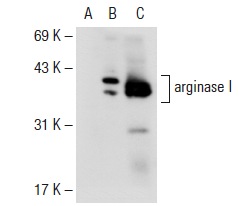
Western blot analysis of arginase I expression in non-transfected: (A) and human arginase I transfected: (B) 293T whole cell lysates and mouse liver tissue extract (C).Application
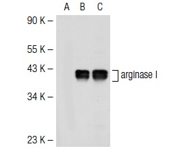
Western blot analysis of arginase I expression in non-transfected: (A) and mouse arginase I transfected: (B) 293T whole cell lysates and mouse liver tissue extract (C).Application
Western blot analysis of arginase I expression in non-transfected: (A) and mouse arginase I transfected: (B) 293T whole cell lysates and mouse liver tissue extract (C).Application
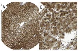
Immunoperoxidase staining of formalin fixed, paraffin-embedded human liver tissue showing nuclear and cytoplasmic staining of hepatocytes at low (A) and high (B) magnification. Kindly provided by The Swedish Human Protein Atlas (HPA) program.Application
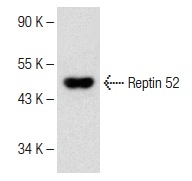
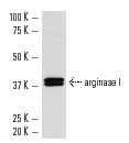
Western blot analysis of arginase I expression in mouse liver tissue extract.| Product Name | arginase I Antibody (H-52) |
|---|---|
| Antibody Type | Primary Antibodies |
| Antigen Alias | epitope corresponding to amino acids 271-322 mapping at the C-terminus of arginase I of human origin |
| Modification Notes | Arginase I (also designated liver-type arginase), which is expressed almost exclusively in the liver, catalyzes the conversion of arginine to ornithine and urea (1). The human arginase I gene, which maps to chromosome 6q23, encodes a 322 amino acid protein. Arginase I exists as a homotrimeric protein and contains a binuclear manganese cluster (2-4). Arginase II catalyzes the same reaction as arginase I, but differs in its tissue specificity and subcellular location (5,6). Specifically, arginase II localizes to the mitochondria (5,6). Arginase II is expressed in non-hepatic tissues, with the highest levels of expression in the kidneys, but, unlike arginase I, is not expressed in liver (5,6). The human arginase II gene, which maps to chromosome 14q24.1-q24.3, encodes a 354 amino acid protein (3,5-7). In addition, arginase II contains a putative amino-terminal mitochondrial localization sequence (5,6). |
| Clonality | Polyclonal |
|---|---|
| Isotype | IgG |
| Host Species | Rabbit |
| Tested Applications | WBIPIFIHCELISA |
| recommended for detection of arginase I of mouse, rat and human origin by WB, IP, IF, IHC(P) and ELISA also reactive with additional species, including equine, canine, bovine and porcine: |
|
| Species Reactivity | HumanMouseRat |
| Concentration | 1mg/ml |
| Purification | Affinity purified |
| Alternative Names | epitope corresponding to amino acids 271-322 mapping at the C-terminus of arginase I of human origin |
|---|---|
| Tissue Specificity | epitope corresponding to amino acids 271-322 mapping at the C-terminus of arginase I of human origin |
| Entrez Gene | 383 |
|---|
Application
Western blot analysis of arginase I expression in non-transfected: (A) and human arginase I transfected: (B) 293T whole cell lysates and mouse liver tissue extract (C).
Application
Western blot analysis of arginase I expression in non-transfected: (A) and mouse arginase I transfected: (B) 293T whole cell lysates and mouse liver tissue extract (C).
Application
Western blot analysis of arginase I expression in non-transfected: (A) and mouse arginase I transfected: (B) 293T whole cell lysates and mouse liver tissue extract (C).
Application
Immunoperoxidase staining of formalin fixed, paraffin-embedded human liver tissue showing nuclear and cytoplasmic staining of hepatocytes at low (A) and high (B) magnification. Kindly provided by The Swedish Human Protein Atlas (HPA) program.
Application
Western blot analysis of arginase I expression in mouse liver tissue extract.| Application Notes | recommended for detection of arginase I of mouse, rat and human origin by WB, IP, IF, IHC(P) and ELISA also reactive with additional species, including equine, canine, bovine and porcine: |
|---|
| Form | Liquid |
|---|---|
| Storage Instructions | For short-term storage, store at 4° C. For long-term storage, aliquot and store at -20ºC or below. Avoid multiple freeze-thaw cycles. |
| Storage Buffer | phosphate buffered saline , pH 7.4, 150mM NaCl, 0.02% sodium azide and 50% glycerol. |


