Application
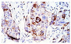
Immunoperoxidase staining of formalin-fixed, paraffin-embedded human breast carcinoma tissue at low magnification.Application
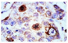
Immunoperoxidase staining of formalin-fixed, paraffin-embedded human breast carcinoma tissue at high magnification. Note staining of selected cells, showing nuclear localization.Application
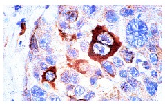
Immunoperoxidase staining of formalin-fixed, paraffin-embedded human breast carcinoma tissue at high magnification. Note staining of selected cells, showing cytoplasmic localization.Application
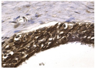
Immunoperoxidase staining of formalin fixed, paraffin-embedded human testis tissue showing nuclear and cytoplasmic staining of cells in seminiferus ducts.Application
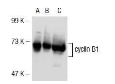
Western blot analysis of cyclin B1 expression in non-transfected: (A) and mouse cyclin B1 transfected: (B) 293T whole cell lysates and K-562 nuclear extract (C).Application
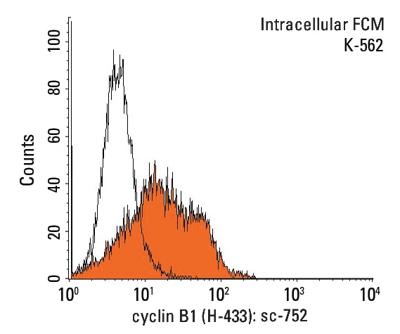
Indirect, intracellular FCM analysis of fixed and permeabilized K-562 cells stained with cyclin B1 (H-433), followed by PE-conjugated goat anti-rabbit IgG: . Black line histogram represents the isotype control, normal rabbit IgG: .Application
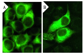
Immunofluorescence staining of methanol-fixed, control HeLa (A) and cyclin B1 siRNA silenced HeLa (B) cells showing diminished cytoplasmic staining in the siRNA silenced cells. Cells probed with cyclin B1 (H-433): .Application
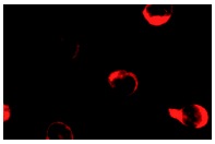
Immunofluorescence staining of methanol-fixed K-562 cells showing cytoplasmic staining.Application
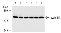
Western blot analysis of cyclin B1 expression in untreated (A,C,E) and phorbol ester-induced (B,D,F) K-562 (A,B), Jurkat (C,D) and HeLa (E,F) nuclear extracts.| Product Name | cyclin B1 Antibody (H-433) TRITC |
|---|---|
| Antibody Type | Primary Antibodies |
| Antigen Alias | epitope corresponding to amino acids 1-433 representing full length cyclin B1 of human origin |
| Modification Notes | In eukaryotic cells, mitosis is initiated following the activation of a protein kinase known variously as maturation-promoting factor, M-phase specific histone kinase or M-phase kinase. This protein kinase is composed of a catalytic subunit (Cdc2), a regulatory subunit (cyclin B) and a low molecular weight subunit (p13-Suc 1). The Cdc/cyclin enzyme is subject to multiple levels of control, of which the regulation of the catalytic subunit by tyrosine phosphorylation is the best understood. Tyrosine phosphorylation inhibits the Cdc2/cyclin B enzyme; tyrosine dephosphorylation, occurring at the onset of mitosis, directly activates the pre-MPF complex. Evidence has established that B type cyclins not only act on M-phase regulatory subunits of the Cdc2 protein kinase, but also activate the Cdc25A and Cdc25B endogenous tyrosine phosphatase, of which Cdc2 is the physiological substrate. The specificity of this effect is shown by the inability of either cyclin A or cyclin D1 to display any such stimulation of Cdc25A or Cdc25B. |
| Clonality | Polyclonal |
|---|---|
| Isotype | IgG |
| Host Species | Rabbit |
| Tested Applications | WBIPIFIHCFCELISA |
| recommended for detection of cyclin B1 of mouse, rat and human origin by WB, IP, IF, IHC(P), ELISA and indirect FCM using PE-conjugated goat anti-rabbit IgG: sc-3739 also reactive with additional species, including equine, canine, bovine and porcine: |
|
| Species Reactivity | HumanMouseRat |
| Concentration | 1mg/ml |
| Purification | Affinity purified |
| Alternative Names | epitope corresponding to amino acids 1-433 representing full length cyclin B1 of human origin |
|---|---|
| Tissue Specificity | epitope corresponding to amino acids 1-433 representing full length cyclin B1 of human origin |
| Entrez Gene | 891 |
|---|
Application
Immunoperoxidase staining of formalin-fixed, paraffin-embedded human breast carcinoma tissue at low magnification.
Application
Immunoperoxidase staining of formalin-fixed, paraffin-embedded human breast carcinoma tissue at high magnification. Note staining of selected cells, showing nuclear localization.
Application
Immunoperoxidase staining of formalin-fixed, paraffin-embedded human breast carcinoma tissue at high magnification. Note staining of selected cells, showing cytoplasmic localization.
Application
Immunoperoxidase staining of formalin fixed, paraffin-embedded human testis tissue showing nuclear and cytoplasmic staining of cells in seminiferus ducts.
Application
Western blot analysis of cyclin B1 expression in non-transfected: (A) and mouse cyclin B1 transfected: (B) 293T whole cell lysates and K-562 nuclear extract (C).
Application
Indirect, intracellular FCM analysis of fixed and permeabilized K-562 cells stained with cyclin B1 (H-433), followed by PE-conjugated goat anti-rabbit IgG: . Black line histogram represents the isotype control, normal rabbit IgG: .
Application
Immunofluorescence staining of methanol-fixed, control HeLa (A) and cyclin B1 siRNA silenced HeLa (B) cells showing diminished cytoplasmic staining in the siRNA silenced cells. Cells probed with cyclin B1 (H-433): .
Application
Immunofluorescence staining of methanol-fixed K-562 cells showing cytoplasmic staining.
Application
Western blot analysis of cyclin B1 expression in untreated (A,C,E) and phorbol ester-induced (B,D,F) K-562 (A,B), Jurkat (C,D) and HeLa (E,F) nuclear extracts.| Application Notes | recommended for detection of cyclin B1 of mouse, rat and human origin by WB, IP, IF, IHC(P), ELISA and indirect FCM using PE-conjugated goat anti-rabbit IgG: sc-3739 also reactive with additional species, including equine, canine, bovine and porcine: |
|---|
| Form | Liquid |
|---|---|
| Storage Instructions | For short-term storage, store at 4° C. For long-term storage, aliquot and store at -20ºC or below. Avoid multiple freeze-thaw cycles. |
| Storage Buffer | phosphate buffered saline , pH 7.4, 150mM NaCl, 0.02% sodium azide and 50% glycerol. |


