Application
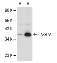
Western blot analysis of AKR7A2 expression in non-transfected: (A) and human AKR7A2 transfected: (B) 293 whole cell lysates.Application
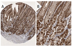
Immunoperoxidase staining of formalin fixed, paraffin-embedded human stomach tissue showing cytoplasmic and nuclear staining of glandular cells in low (A) and high (B) resolution. Kindly provided by The Swedish Human Protein Atlas (HPA) program.Application
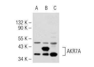
Western blot analysis of AKR7A expression in non-transfected 293T: (A), human AKR7 transfected 293T: (B) and Hep G2 (C) whole cell lysates.Application
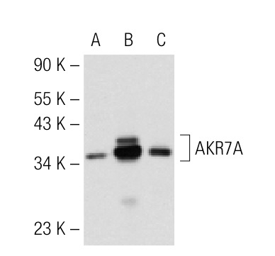
Western blot analysis of AKR7 Aexpression in non-transfected 293T: (A), human AKR7 transfected 293T: (B) and HL-60 (C) whole cell lysates.Application
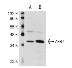
Western blot analysis of AKR7 expression in 293T (A) and HL-60 (B) whole cell lysates.Application
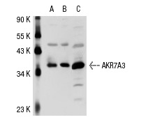
Western blot analysis of AKR7A3 expression in non-transfected 293T: (A), human AKR7A3 transfected 293T: (B) and HeLa (C) whole cell lysates.Application
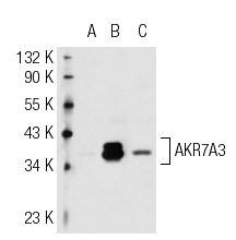
Western blot analysis of AKR7A3 expression in non-transfected 293T: (A), human AKR7A3 transfected 293T: (B) and Hep G2 (C) whole cell lysates.| Product Name | AKR7A Antibody (H-300) |
|---|---|
| Antibody Type | Primary Antibodies |
| Antigen Alias | epitope corresponding to amino acids 63-359 mapping at the C-terminus of AKR7A2 of human origin |
| Modification Notes | The aldo-keto reductase 7 (AKR7) family includes AKR7A2, AKR7A3 and AKR7A4 in human, AKR7A5 in mouse and AKR7A2 in rat, all of which function in the metabolism of Aflatoxin B1 and other dicarbonyl-containing compounds. More specifically, AKR7A proteins are involved in the metabolism of compounds with ketone groups on adjacent carbon atoms in a broad range of tissues, notably the liver. The human AKR7A2 gene maps to human chromosome 1p36.13, a region frequently deleted in sporadic colorectal cancer. The functional significance of this correlation lies in the constitutive expression of AKR7A2 in human liver to eliminate Aflatoxin (an environmental carcinogen), thus acting as an endogenous chemo-preventative agent. AKR7A3 is believed to be a homodimer expressed in kidney, colon, pancreas, endometrium and adenocarcinoma. |
| Clonality | Polyclonal |
|---|---|
| Isotype | IgG |
| Host Species | Rabbit |
| Tested Applications | WBIPIFIHCELISA |
| recommended for detection of AKR7A2, AKR7A3 and AKR7A4 of human origin, AKR7A5 of mouse origin and AKR7A2 of rat origin by WB, IP, IF, IHC(P) and ELISA also reactive with additional species, including equine, canine, bovine and porcine: |
|
| Species Reactivity | HumanMouseRat |
| Concentration | 1mg/ml |
| Purification | Affinity purified |
| Alternative Names | epitope corresponding to amino acids 63-359 mapping at the C-terminus of AKR7A2 of human origin |
|---|---|
| Tissue Specificity | epitope corresponding to amino acids 63-359 mapping at the C-terminus of AKR7A2 of human origin |
| Entrez Gene | 8574 |
|---|
Application
Western blot analysis of AKR7A2 expression in non-transfected: (A) and human AKR7A2 transfected: (B) 293 whole cell lysates.
Application
Immunoperoxidase staining of formalin fixed, paraffin-embedded human stomach tissue showing cytoplasmic and nuclear staining of glandular cells in low (A) and high (B) resolution. Kindly provided by The Swedish Human Protein Atlas (HPA) program.
Application
Western blot analysis of AKR7A expression in non-transfected 293T: (A), human AKR7 transfected 293T: (B) and Hep G2 (C) whole cell lysates.
Application
Western blot analysis of AKR7 Aexpression in non-transfected 293T: (A), human AKR7 transfected 293T: (B) and HL-60 (C) whole cell lysates.
Application
Western blot analysis of AKR7 expression in 293T (A) and HL-60 (B) whole cell lysates.
Application
Western blot analysis of AKR7A3 expression in non-transfected 293T: (A), human AKR7A3 transfected 293T: (B) and HeLa (C) whole cell lysates.
Application
Western blot analysis of AKR7A3 expression in non-transfected 293T: (A), human AKR7A3 transfected 293T: (B) and Hep G2 (C) whole cell lysates.| Application Notes | recommended for detection of AKR7A2, AKR7A3 and AKR7A4 of human origin, AKR7A5 of mouse origin and AKR7A2 of rat origin by WB, IP, IF, IHC(P) and ELISA also reactive with additional species, including equine, canine, bovine and porcine: |
|---|
| Form | Liquid |
|---|---|
| Storage Instructions | For short-term storage, store at 4° C. For long-term storage, aliquot and store at -20ºC or below. Avoid multiple freeze-thaw cycles. |
| Storage Buffer | phosphate buffered saline , pH 7.4, 150mM NaCl, 0.02% sodium azide and 50% glycerol. |


