IF
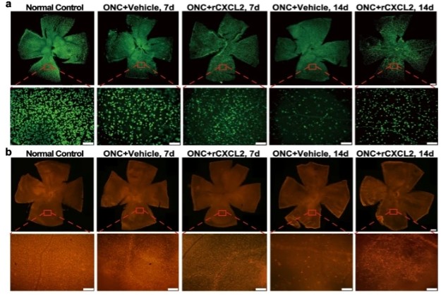
a, b Immunofluorescence staining images showing RBPMS- or Thy1-positive RGCs in the whole-mount retinas (scale bar: 200 μm) and their representative images of middle regions in the retina (scale bar: 50 μm) 7 and 14 days after ONC with vehicle (PBS) or rCXCL2 injection, respectively. The whole retina was photomerged by several images at low magnification using Adobe Photoshop software.IF
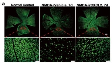
a RBPMS staining images of whole-mount retinas (scale bar: 200 μm) and representative images of middle regions in the retina (scale bar: 50 μm) for different groups. The whole retina was photomerged by several images at low magnification with Adobe Photoshop software.IF
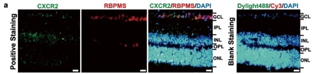
a Immunofluorescence images of retinal sections to visualize the specific CXCL2 receptor, CXCR2 (green), RGCs (anti-RBPMS) (red), and cellular nucleus (DAPI) (blue). Scale bar, 20 μm.| Product Name | RBPMS Antibody (N-20) |
|---|---|
| Antibody Type | Primary Antibodies |
| Modification Notes | RBPMS (RNA binding protein with multiple splicing), also known as HERMES, is a 196 amino acid protein that contains one RRM (RNA recognition motif) domain and belongs to the RRM family of RNA-binding proteins. Expressed ubiquitously, RBPMS exists as multiple alternatively spliced isoforms and is thought to bind RNA, possibly playing a role in RNA-related events, such as transcription and translation. The gene encoding RBPMS maps to human chromosome 8, which consists of nearly 146 million base pairs, houses more than 800 genes and is associated with a variety of diseases and malignancies. Schizophrenia, bipolar disorder, Trisomy 8, Pfeiffer syndrome, congenital hypothyroidism, Waardenburg syndrome and some leukemias and lymphomas are thought to occur as a result of defects in specific genes that map to chromosome 8. |
| Clonality | Polyclonal |
|---|---|
| Isotype | IgG |
| Host Species | Rabbit |
| Tested Applications | ELISAIFWB |
| recommended for detection of RBPMS of mouse, rat and human origin by WB, IF and ELISA also reactive with additional species, including equine, canine, bovine and porcine: |
|
| Species Reactivity | HumanMouseRat |
| Concentration | 1mg/ml |
| Purification | Affinity purified |
| Alternative Names | epitope mapping at the N-terminus of RBPMS of human origin |
|---|---|
| Tissue Specificity | epitope mapping at the N-terminus of RBPMS of human origin |
| Entrez Gene | 11030 |
|---|
IF
a, b Immunofluorescence staining images showing RBPMS- or Thy1-positive RGCs in the whole-mount retinas (scale bar: 200 μm) and their representative images of middle regions in the retina (scale bar: 50 μm) 7 and 14 days after ONC with vehicle (PBS) or rCXCL2 injection, respectively. The whole retina was photomerged by several images at low magnification using Adobe Photoshop software.
IF
a RBPMS staining images of whole-mount retinas (scale bar: 200 μm) and representative images of middle regions in the retina (scale bar: 50 μm) for different groups. The whole retina was photomerged by several images at low magnification with Adobe Photoshop software.
IF
a Immunofluorescence images of retinal sections to visualize the specific CXCL2 receptor, CXCR2 (green), RGCs (anti-RBPMS) (red), and cellular nucleus (DAPI) (blue). Scale bar, 20 μm.| Application Notes | recommended for detection of RBPMS of mouse, rat and human origin by WB, IF and ELISA also reactive with additional species, including equine, canine, bovine and porcine: |
|---|
| Form | Liquid |
|---|---|
| Storage Instructions | For short-term storage, store at 4° C. For long-term storage, aliquot and store at -20ºC or below. Avoid multiple freeze-thaw cycles. |
| Storage Buffer | phosphate buffered saline , pH 7.4, 150mM NaCl, 0.02% sodium azide and 50% glycerol. |


