Application
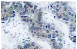
Immunoperoxidase staining of formalin-fixed, paraffin-embedded human breast carcinoma tissue showing membrane and cytoplasmic localization.WB
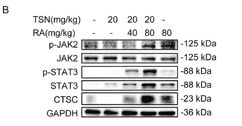
(B) The expressions of p-JAK2 (Tyr1007), JAK2, p-STAT3 (Tyr705), STAT3 and CTSC in the liver were analyzed by western blot.WB
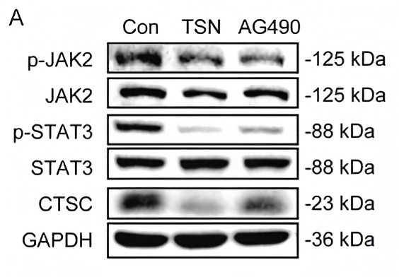
(A) HepG2 cells treated with 10 μM TSN or 50 μM AG490 for 24 h were analyzed by western blot for the protein levels of p-JAK2 (Tyr1007), JAK2, p-STAT3 (Tyr705), STAT3 and CTSC.WB
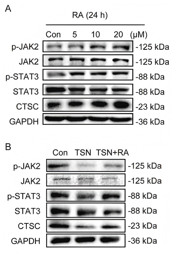
(A) HepG2 cells treated with RA (0, 5, 10, 20 μM) for 24 h were analyzed by western blot for the protein levels of p-JAK2 (Tyr1007), JAK2, p-STAT3 (Tyr705), STAT3 and CTSC. (B) HepG2 cells were treated with 10 μM TSN for 24 h after pretreatment with 20 μM RA or vehicle for 3 h. The expressions of p-JAK2 (Tyr1007), JAK2, p-STAT3 (Tyr705), STAT3 and CTSC were analyzed by western blot. HepG2 cells were treated by 10 μM TSN or 20 μM RA for 24 h with 20 μM RA pretreatment for 3 h.WB
(A-B) The expression levels of p-JAK2 (Tyr1007), JAK2, p-STAT3 (Tyr705) and STAT3 were analyzed by Western blot.WB
(B) The protein expressions of p-JAK2 (Tyr1007), JAK2, p-STAT3 (Tyr705), STAT3, LC3B and P62 were analyzed by Western blot.WB
| Product Name | p-JAK2 Antibody (Tyr 1007) |
|---|---|
| Antibody Type | Primary Antibodies |
| Modification | Phosphoralated |
| Modification Notes | JAK2 (Janus Kinase 2) belongs to the emerging family of non-receptor Janus tyrosine kinases, which regulate a spectrum of cellular functions downstream of activated cytokine receptors in the lympho-hematopoietic system. Immuno-logical stimuli, such as interferons and cytokines, induce recruitment of Stat transcription factors to cytokine receptor-associated JAK2. JAK2 then phosphorylates proximal Stat factors, which subsequently dimerize, translocate to the nucleus and bind to cis elements upstream of target gene promoters to regulate transcription. The canonical JAK-Stat pathway is integral to maintaining a normal immune system by stimulating proliferation, differentiation, survival, and host resistance to pathogens. Altering JAK-Stat signaling to reduce cytokine induced pro-inflammatory responses represents an attractive target for anti-inflammatory therapies. Within the JAK2 kinase domain, there is a region that has considerable sequence homology to the regulatory region of the insulin receptor. Among a variety of sites, Tyrosines 1007 and 1008 are sites of trans- or autophosphorylation in vivo and in in vitro kinase reactions.. |
| Clonality | Polyclonal |
|---|---|
| Isotype | IgG |
| Host Species | Rabbit |
| Tested Applications | IFIHCWB |
| recommended for detection of Tyr 1007 phosphorylated JAK2 of mouse, rat and human origin by IF and IHC(P): |
|
| Species Reactivity | HumanMouseRat |
| Concentration | 1mg/ml |
| Purification | Affinity purified |
| Alternative Names | epitope corresponding to phosphorylated Tyr 1007 of JAK2 of human origin |
|---|---|
| Tissue Specificity | epitope corresponding to phosphorylated Tyr 1007 of JAK2 of human origin |
| Entrez Gene | 3717 |
|---|
Application
Immunoperoxidase staining of formalin-fixed, paraffin-embedded human breast carcinoma tissue showing membrane and cytoplasmic localization.
WB
(B) The expressions of p-JAK2 (Tyr1007), JAK2, p-STAT3 (Tyr705), STAT3 and CTSC in the liver were analyzed by western blot.
WB
(A) HepG2 cells treated with 10 μM TSN or 50 μM AG490 for 24 h were analyzed by western blot for the protein levels of p-JAK2 (Tyr1007), JAK2, p-STAT3 (Tyr705), STAT3 and CTSC.
WB
(A) HepG2 cells treated with RA (0, 5, 10, 20 μM) for 24 h were analyzed by western blot for the protein levels of p-JAK2 (Tyr1007), JAK2, p-STAT3 (Tyr705), STAT3 and CTSC. (B) HepG2 cells were treated with 10 μM TSN for 24 h after pretreatment with 20 μM RA or vehicle for 3 h. The expressions of p-JAK2 (Tyr1007), JAK2, p-STAT3 (Tyr705), STAT3 and CTSC were analyzed by western blot. HepG2 cells were treated by 10 μM TSN or 20 μM RA for 24 h with 20 μM RA pretreatment for 3 h.
WB
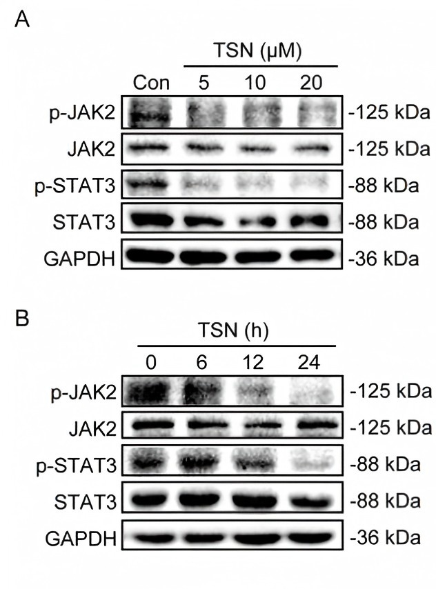
(A-B) The expression levels of p-JAK2 (Tyr1007), JAK2, p-STAT3 (Tyr705) and STAT3 were analyzed by Western blot.
WB
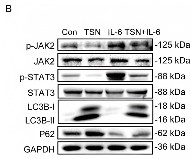
(B) The protein expressions of p-JAK2 (Tyr1007), JAK2, p-STAT3 (Tyr705), STAT3, LC3B and P62 were analyzed by Western blot.WB
| Application Notes | recommended for detection of Tyr 1007 phosphorylated JAK2 of mouse, rat and human origin by IF and IHC(P): |
|---|
| Form | Liquid |
|---|---|
| Storage Instructions | For short-term storage, store at 4° C. For long-term storage, aliquot and store at -20ºC or below. Avoid multiple freeze-thaw cycles. |
| Storage Buffer | phosphate buffered saline , pH 7.4, 150mM NaCl, 0.02% sodium azide and 50% glycerol. |


