Application
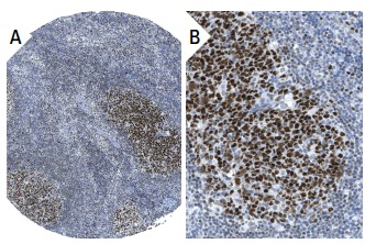
Immunoperoxidase staining of formalin fixed, paraffin-embedded human lymph node tissue showing nuclear staining of reaction center cells at low (A) and high (B) magnification. Kindly provided by The Swedish Human Protein Atlas (HPA) program.Application
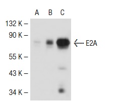
Western blot analysis of E2A expression in non-transfected 293T: (A), mouse E2A transfected 293T: (B) and Jurkat (C) whole cell lysates.Application
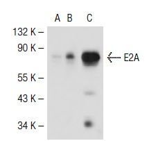
Western blot analysis of E2A expression in non-transfected 293T: (A), mouse E2A transfected 293T: (B) and Jurkat (C) whole cell lysates.Application
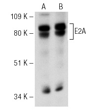
Western blot analysis of E2A expression in non-transfected: (A) and mouse E2A transfected: (B) 293 whole cell lysates.Application
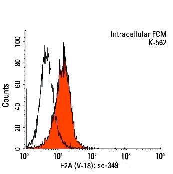
Indirect, intracellular FCM analysis of fixed and permeabilized K-562 cells stained with E2A (V-18), followed by PE-conjugated goat anti-rabbit IgG: . Black line histogram represents the isotype control, normal rabbit IgG: .Application
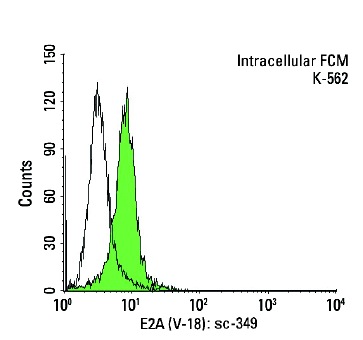
Indirect, intracellular FCM analysis of fixed and permeabilized K-562 cells stained with E2A (V-18), followed by FITC-conjugated goat anti-rabbit IgG: . Black line histogram represents the isotype control, normal rabbit IgG: .Application
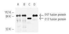
Antibodies tested include E2A (N-649): (A), E2A (Yae): (B) and E2A (V-18): (C,D).Application
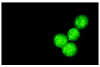
Immunofluorescence staining of methanol-fixed K-562 cells showing nuclear staining.Application
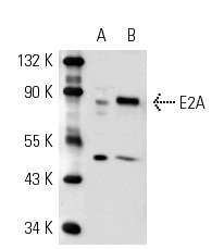
Western blot analysis of E2A expression in Jurkat (A) and K-562 (B) whole cell lysates.| Product Name | E2A Antibody (V-18) |
|---|---|
| Antibody Type | Primary Antibodies |
| Antigen Alias | epitope mapping at the C-terminus of E2A of human origin |
| Modification Notes | Differentiation of myogenic cells is regulated by multiple positively and negatively acting factors. One well characterized family of helix-loop-helix (HLH) proteins known to play an important role in the regulation of muscle cell development includes MyoD, myogenin, Myf-5 and Myf-6 (also designated MRF-4 or herculin). MyoD transcription factors form heterodimers with products of a more widely expressed family of bHLH genes, the E family, which consists of at least three distinct genes: E2A, IF2 and HEB. MyoD-E heterodimers bind avidly to consensus (CANNTG) E box target sites that are functionally important elements in the upstream regulatory sequences of many muscle-specific terminal differentiation genes. Both homo- and hetero-oligomers of these proteins are able to distinguish very closely related E box proteins and are believed to play important roles in lineage specific gene expression. |
| Clonality | Polyclonal |
|---|---|
| Isotype | IgG |
| Host Species | Rabbit |
| Tested Applications | WBIPIFIHCFCELISAGSChIP |
| recommended for detection of E2A isoforms E12 and E47 of mouse, rat and human origin by WB, IP, IF, IHC(P), ELISA and indirect FCM using PE (sc-3739)- or FITC (sc-2012)-conjugated goat anti-rabbit IgG also reactive with additional species, including equine, canine and bovine: |
|
| Species Reactivity | HumanMouseRat |
| Concentration | 1mg/ml |
| Purification | Affinity purified |
| Alternative Names | epitope mapping at the C-terminus of E2A of human origin |
|---|---|
| Tissue Specificity | epitope mapping at the C-terminus of E2A of human origin |
| Entrez Gene | 6929 |
|---|
Application
Immunoperoxidase staining of formalin fixed, paraffin-embedded human lymph node tissue showing nuclear staining of reaction center cells at low (A) and high (B) magnification. Kindly provided by The Swedish Human Protein Atlas (HPA) program.
Application
Western blot analysis of E2A expression in non-transfected 293T: (A), mouse E2A transfected 293T: (B) and Jurkat (C) whole cell lysates.
Application
Western blot analysis of E2A expression in non-transfected 293T: (A), mouse E2A transfected 293T: (B) and Jurkat (C) whole cell lysates.
Application
Western blot analysis of E2A expression in non-transfected: (A) and mouse E2A transfected: (B) 293 whole cell lysates.
Application
Indirect, intracellular FCM analysis of fixed and permeabilized K-562 cells stained with E2A (V-18), followed by PE-conjugated goat anti-rabbit IgG: . Black line histogram represents the isotype control, normal rabbit IgG: .
Application
Indirect, intracellular FCM analysis of fixed and permeabilized K-562 cells stained with E2A (V-18), followed by FITC-conjugated goat anti-rabbit IgG: . Black line histogram represents the isotype control, normal rabbit IgG: .
Application
Antibodies tested include E2A (N-649): (A), E2A (Yae): (B) and E2A (V-18): (C,D).
Application
Immunofluorescence staining of methanol-fixed K-562 cells showing nuclear staining.
Application
Western blot analysis of E2A expression in Jurkat (A) and K-562 (B) whole cell lysates.| Application Notes | recommended for detection of E2A isoforms E12 and E47 of mouse, rat and human origin by WB, IP, IF, IHC(P), ELISA and indirect FCM using PE (sc-3739)- or FITC (sc-2012)-conjugated goat anti-rabbit IgG also reactive with additional species, including equine, canine and bovine: |
|---|
| Form | Liquid |
|---|---|
| Storage Instructions | For short-term storage, store at 4° C. For long-term storage, aliquot and store at -20ºC or below. Avoid multiple freeze-thaw cycles. |
| Storage Buffer | phosphate buffered saline , pH 7.4, 150mM NaCl, 0.02% sodium azide and 50% glycerol. |


