Application
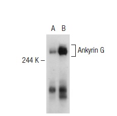
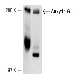
Western blot analysis of Ankyrin G expression in non-transfected: (A) and human Ankyrin G transfected: (B) 293T whole cell lysates.Application
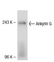
Western blot analysis of Ankyrin G expression in rat brain tissue extract.Application
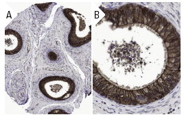
Immunoperoxidase staining of formalin fixed, paraffin-embedded human epididymus tissue showing cytoplasmic, membrane, and nuclear staining of glandular cells at low (A) and high (B) magnification. Kindly provided by The Swedish Human Protein Atlas (HPA) program.Application
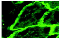
Immunofluorescence staining of normal mouse skin frozen section showing membrane staining.Application
Western blot analysis of Ankyrin G expression in Caco-2 (A) and IMR-32 (B) whole cell lysates.WB
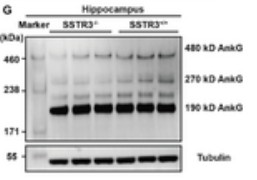
G. Western blotting of AnkG expression of hippocampus in SSTR3+/+ and SSTR3−/− mice, using β-tubulin as a reference.IF
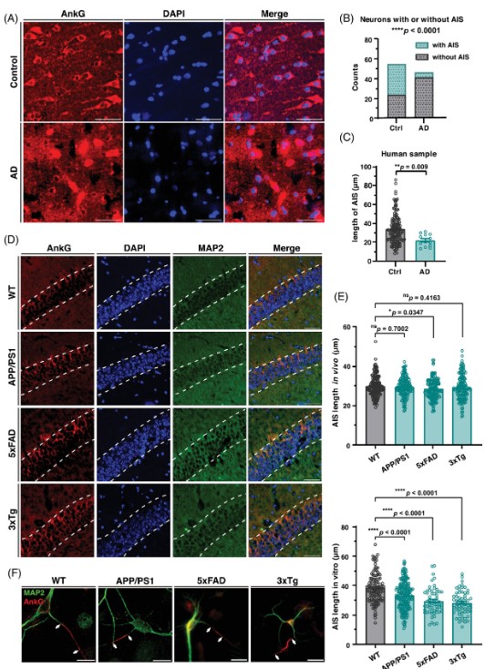
Neurons in vivo and in vitro from Alzheimer's disease (AD) model mice and human brain samples have altered axon initial segment (AIS) lengths. (A) Immunofluorescence staining of paraffin-sectioned samples of human cerebral cortex. Red, AnkG (AIS); blue, DAPI (nuclei). Scale bar, 50 µm. (B) Statistics of staining results in (A). The number of neurons clearly visible with or without AIS was counted in the two samples. Data analyzed by Fisher's exact test. (C) Statistics performed on the lengths of the AIS in brain slices from human samples. Student's t-test was used for analysis. (D) Immunofluorescence staining in coronal frozen sections at the CA1 region of the hippocampus. Slices were from 6-month-old wild-type, APP/PS1, 5xFAD, and 3xTg mice. The dashed line is the outline of the cluster of cell bodies in the CA1 region of the hippocampus. Red, AnkG (AIS), green, MAP2 (soma and dendrites), blue, DAPI (nuclei). Scale bar, 50 µm. (F) Immunofluorescence staining of DIV 7 cultured neurons from wild-type, APP/PS1, 5xFAD, and 3xTg E18.5 mice. The segment between the two arrow marks is the AIS identified by AnkG. Red, AnkG, green, MAP2. Scale bar, 20 µm. (E and G) Statistics performed on the lengths of the AIS in brain slices and cultured neurons from the four strains. The upper panel shows the results of brain slices, and the lower panel shows the results of cultured neurons. One-way analysis of variance (ANOVA) was used for analysis. ns, not significant; *p < 0.05; ****p < 0.0001.| Product Name | Ankyrin G Antibody (H-215) |
|---|---|
| Antibody Type | Primary Antibodies |
| Modification Notes | Members of the Ankyrin family of proteins mediate the attachment of integral membrane proteins to the cytoskeleton. ANK1, ANK2 and ANK3 genes encode for the proteins in this family, Ankyrin-1 (also designated Ankyrin R), Ankyrin B and Ankyrin G, respectively. The proteins are structured similarly, each composed of an N-terminal domain with multiple ankyrin repeats, a highly conserved central spectrin binding domain and C-terminal regulatory domains which are susceptible to the most variance. Both Ankyrin B and Ankyrin G are essential for normal neuronal functions. Ankyrin G was originally identified localized to the nodes of Ranvier and axon initial segments where it was found to mediate normal neuronal sodium channel function. Ankyrin G, named for its giant size and general expression, exhibits tissue-specific alternative mRNA processing. The two largest protein isoforms are expressed only in nervous tissue, with additional smaller isoforms found in an array of other tissues including lung, kidney, spleen, liver and heart. |
| Clonality | Polyclonal |
|---|---|
| Isotype | IgG |
| Host Species | Rabbit |
| Tested Applications | ELISAIFIHCIPWB |
| recommended for detection of Ankyrin G of mouse, rat and human origin by WB, IP, IF, IHC(P) and ELISA also reactive with additional species, including equine, canine and porcine: |
|
| Species Reactivity | HumanMouseRat |
| Concentration | 1mg/ml |
| Purification | Affinity purified |
| Alternative Names | epitope corresponding to amino acids 4163-4377 mapping at the C-terminus of Ankyrin G of human origin |
|---|---|
| Tissue Specificity | epitope corresponding to amino acids 4163-4377 mapping at the C-terminus of Ankyrin G of human origin |
| Entrez Gene | 288 |
|---|
Application
Western blot analysis of Ankyrin G expression in non-transfected: (A) and human Ankyrin G transfected: (B) 293T whole cell lysates.
Application
Western blot analysis of Ankyrin G expression in rat brain tissue extract.
Application
Immunoperoxidase staining of formalin fixed, paraffin-embedded human epididymus tissue showing cytoplasmic, membrane, and nuclear staining of glandular cells at low (A) and high (B) magnification. Kindly provided by The Swedish Human Protein Atlas (HPA) program.
Application
Immunofluorescence staining of normal mouse skin frozen section showing membrane staining.
Application
Western blot analysis of Ankyrin G expression in Caco-2 (A) and IMR-32 (B) whole cell lysates.
WB
G. Western blotting of AnkG expression of hippocampus in SSTR3+/+ and SSTR3−/− mice, using β-tubulin as a reference.
IF
Neurons in vivo and in vitro from Alzheimer's disease (AD) model mice and human brain samples have altered axon initial segment (AIS) lengths. (A) Immunofluorescence staining of paraffin-sectioned samples of human cerebral cortex. Red, AnkG (AIS); blue, DAPI (nuclei). Scale bar, 50 µm. (B) Statistics of staining results in (A). The number of neurons clearly visible with or without AIS was counted in the two samples. Data analyzed by Fisher's exact test. (C) Statistics performed on the lengths of the AIS in brain slices from human samples. Student's t-test was used for analysis. (D) Immunofluorescence staining in coronal frozen sections at the CA1 region of the hippocampus. Slices were from 6-month-old wild-type, APP/PS1, 5xFAD, and 3xTg mice. The dashed line is the outline of the cluster of cell bodies in the CA1 region of the hippocampus. Red, AnkG (AIS), green, MAP2 (soma and dendrites), blue, DAPI (nuclei). Scale bar, 50 µm. (F) Immunofluorescence staining of DIV 7 cultured neurons from wild-type, APP/PS1, 5xFAD, and 3xTg E18.5 mice. The segment between the two arrow marks is the AIS identified by AnkG. Red, AnkG, green, MAP2. Scale bar, 20 µm. (E and G) Statistics performed on the lengths of the AIS in brain slices and cultured neurons from the four strains. The upper panel shows the results of brain slices, and the lower panel shows the results of cultured neurons. One-way analysis of variance (ANOVA) was used for analysis. ns, not significant; *p < 0.05; ****p < 0.0001.| Application Notes | recommended for detection of Ankyrin G of mouse, rat and human origin by WB, IP, IF, IHC(P) and ELISA also reactive with additional species, including equine, canine and porcine: |
|---|
| Form | Liquid |
|---|---|
| Storage Instructions | For short-term storage, store at 4° C. For long-term storage, aliquot and store at -20ºC or below. Avoid multiple freeze-thaw cycles. |
| Storage Buffer | phosphate buffered saline , pH 7.4, 150mM NaCl, 0.02% sodium azide and 50% glycerol. |


