Application
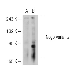
Western blot analysis of Nogo expression in non-transfected: (A) and human Nogo transfected: (B) 293T whole cell lysates.Application
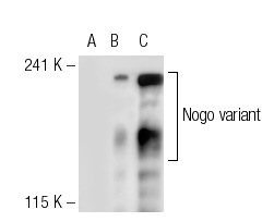
Western blot analysis of Nogo expression in non-transfected 293T: (A), human Nogo transfected 293T: (B) and TE 671 (C) whole cell lysates.Application
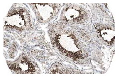
Immunoperoxidase staining of formalin fixed, paraffin-embedded human testis tissue showing cytoplasmic staining of cells in ductus seminiferus. Kindly provided by The Swedish Human Protein Atlas (HPA) program.Application
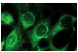
Immunoperoxidase staining of formalin fixed, paraffin-embedded mouse brain tissue showing cytoplasmic localization.Application
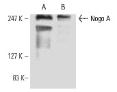
Western blot analysis of Nogo A expression in rat brain tissue extract (A) and F9 whole cell lysate (B).| Product Name | Nogo A Antibody (H-300) |
|---|---|
| Antibody Type | Primary Antibodies |
| Antigen Alias | epitope corresponding to amino acids 700-1000 mapping to an internal region of Nogo A of human origin |
| Modification Notes | CNS white matter is selectively inhibitory for axonal out-growth (1-3). Nogo (also designated NI250 and Reticulon 4-A) is an oligodendrocyte-specific member of the Reticulon family and is a component of CNS white matter that inhibits axon outgrowth, induces collapse of growth cones of chick dorsal root ganglion cells, and inhibits the spreading of 3T3 fibroblasts (4,5). Other members of the reticulin protein family do not inhibit axon extension and are thought to have a role in ER function (4,6). Nogo is expressed by oligodendrocytes but not by Schwann cells, and associates primarily with the endoplasmic reticulum (4). Nogo exists in three different splice forms, Nogo-A, -B and -C (4,7,8). |
| Clonality | Polyclonal |
|---|---|
| Isotype | IgG |
| Host Species | Rabbit |
| Tested Applications | WBIPIFIHCELISA |
| recommended for detection of Nogo A of mouse, rat and human origin by WB, IP, IF, IHC(P) and ELISA: |
|
| Species Reactivity | HumanMouseRat |
| Concentration | 1mg/ml |
| Purification | Affinity purified |
| Alternative Names | epitope corresponding to amino acids 700-1000 mapping to an internal region of Nogo A of human origin |
|---|---|
| Tissue Specificity | epitope corresponding to amino acids 700-1000 mapping to an internal region of Nogo A of human origin |
| Entrez Gene | 57142 |
|---|
Application
Western blot analysis of Nogo expression in non-transfected: (A) and human Nogo transfected: (B) 293T whole cell lysates.
Application
Western blot analysis of Nogo expression in non-transfected 293T: (A), human Nogo transfected 293T: (B) and TE 671 (C) whole cell lysates.
Application
Immunoperoxidase staining of formalin fixed, paraffin-embedded human testis tissue showing cytoplasmic staining of cells in ductus seminiferus. Kindly provided by The Swedish Human Protein Atlas (HPA) program.
Application
Immunoperoxidase staining of formalin fixed, paraffin-embedded mouse brain tissue showing cytoplasmic localization.
Application
Western blot analysis of Nogo A expression in rat brain tissue extract (A) and F9 whole cell lysate (B).| Application Notes | recommended for detection of Nogo A of mouse, rat and human origin by WB, IP, IF, IHC(P) and ELISA: |
|---|
| Form | Liquid |
|---|---|
| Storage Instructions | For short-term storage, store at 4° C. For long-term storage, aliquot and store at -20ºC or below. Avoid multiple freeze-thaw cycles. |
| Storage Buffer | phosphate buffered saline , pH 7.4, 150mM NaCl, 0.02% sodium azide and 50% glycerol. |


