Application
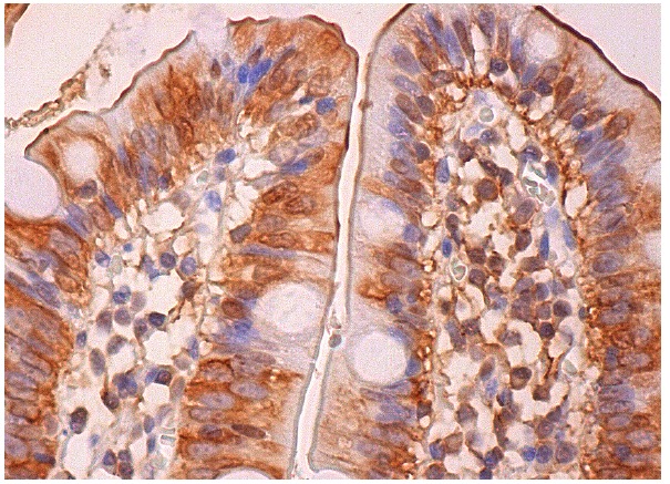
Immunoperox-idase staining of formalin fixed, paraffin-embedded human small intestine tissue showing cytoplasmic, membrane and nuclear staining of glandular cells.Application
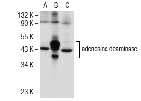
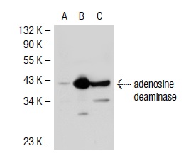
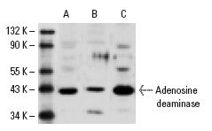
Western blot analysis of adenosine deaminase expression in non-transfected: (A) and human adenosine deaminase transfected: (B) 293T whole cell lysates and rat spleen tissue extract (C).Application
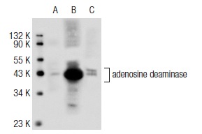
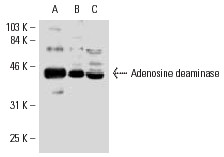
Western blot analysis of adenosine deaminase expression in non-transfected 293T: (A), human adenosine deaminase transfected 293T: (B) and Jurkat (C) whole cell lysates.Application
Western blot analysis of adenosine deaminase expression in non-transfected 293T: (A), human adenosine deaminase transfected 293T: (B) and Jurkat (C) whole cell lysates.Application
Western blot analysis of adenosine deaminase expression in non-transfected 293T: (A), mouse adenosine deaminase transfected 293T: (B) and Jurkat (C) whole cell lysates.Application
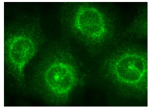
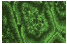
Immunofluorescence staining of normal mouse intestine frozen section showing cytoplasmic staining.Application
Western blot analysis of Adenosine deaminase expression in Jurkat (A) and MM-142 (B) whole cell lysates and rat spleen tissue extract (C).Application
Western blot analysis of Adenosine deaminase expression in Jurkat (A) and MM-142 (B) whole cell lysates.Application
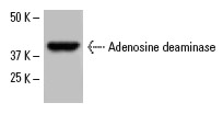
Western blot analysis of Adenosine deaminase expression in Jurkat whole cell lysate.Application
Western blot analysis of Adenosine deaminase expression in NFS-5 C-1 (A), IB4 (B) and MM-142 (C) whole cell lysates.Application
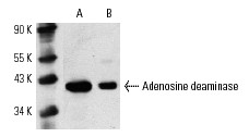
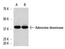
Western blot analysis of Adenosine deaminase expression in Jurkat (A) and MOLT-4 (B) whole cell lysates.| Product Name | adenosine deaminase Antibody (H-300) |
|---|---|
| Antibody Type | Primary Antibodies |
| Antigen Alias | epitope corresponding to amino acids 64-363 mapping at the C-terminus of adensoine deaminase of human origin |
| Modification Notes | Adenosine deaminase is an enzyme that is present in most tissues. It exists predominantly as a monomer, although in some tissues it is associated with adenosine deaminase-binding protein (1). Adenosine deaminase degrades extracellular adenosine, which is toxic for lymphocytes (2). Adenosine deaminase also effects co-stimulatory signals in T cells via interactions with CD26 (3,4). Deficiency of adenosine deaminase has been shown to lead to immunodeficiency diseases such as SCID (severe combined immunodeficiency disease) and has been associated with hereditary hemolytic anemia, a disease in which adenosine deaminase levels are elevated fifty to seventy fold (5,6). |
| Clonality | Polyclonal |
|---|---|
| Isotype | IgG |
| Host Species | Rabbit |
| Tested Applications | WBIPIFIHCELISA |
| recommended for detection of adensoine deaminase of mouse, rat and human origin by WB, IP, IF, IHC(P) and ELISA also reactive with additional species, including equine, canine and bovine: |
|
| Species Reactivity | HumanMouseRat |
| Concentration | 1mg/ml |
| Purification | Affinity purified |
| Alternative Names | epitope corresponding to amino acids 64-363 mapping at the C-terminus of adensoine deaminase of human origin |
|---|---|
| Tissue Specificity | epitope corresponding to amino acids 64-363 mapping at the C-terminus of adensoine deaminase of human origin |
| Entrez Gene | 100 |
|---|
Application
Immunoperox-idase staining of formalin fixed, paraffin-embedded human small intestine tissue showing cytoplasmic, membrane and nuclear staining of glandular cells.
Application
Western blot analysis of adenosine deaminase expression in non-transfected: (A) and human adenosine deaminase transfected: (B) 293T whole cell lysates and rat spleen tissue extract (C).
Application
Western blot analysis of adenosine deaminase expression in non-transfected 293T: (A), human adenosine deaminase transfected 293T: (B) and Jurkat (C) whole cell lysates.
Application
Western blot analysis of adenosine deaminase expression in non-transfected 293T: (A), human adenosine deaminase transfected 293T: (B) and Jurkat (C) whole cell lysates.
Application
Western blot analysis of adenosine deaminase expression in non-transfected 293T: (A), mouse adenosine deaminase transfected 293T: (B) and Jurkat (C) whole cell lysates.
Application
Immunofluorescence staining of normal mouse intestine frozen section showing cytoplasmic staining.
Application
Western blot analysis of Adenosine deaminase expression in Jurkat (A) and MM-142 (B) whole cell lysates and rat spleen tissue extract (C).
Application
Western blot analysis of Adenosine deaminase expression in Jurkat (A) and MM-142 (B) whole cell lysates.
Application
Western blot analysis of Adenosine deaminase expression in Jurkat whole cell lysate.
Application
Western blot analysis of Adenosine deaminase expression in NFS-5 C-1 (A), IB4 (B) and MM-142 (C) whole cell lysates.
Application
Western blot analysis of Adenosine deaminase expression in Jurkat (A) and MOLT-4 (B) whole cell lysates.| Application Notes | recommended for detection of adensoine deaminase of mouse, rat and human origin by WB, IP, IF, IHC(P) and ELISA also reactive with additional species, including equine, canine and bovine: |
|---|
| Form | Liquid |
|---|---|
| Storage Instructions | For short-term storage, store at 4° C. For long-term storage, aliquot and store at -20ºC or below. Avoid multiple freeze-thaw cycles. |
| Storage Buffer | phosphate buffered saline , pH 7.4, 150mM NaCl, 0.02% sodium azide and 50% glycerol. |


