Application
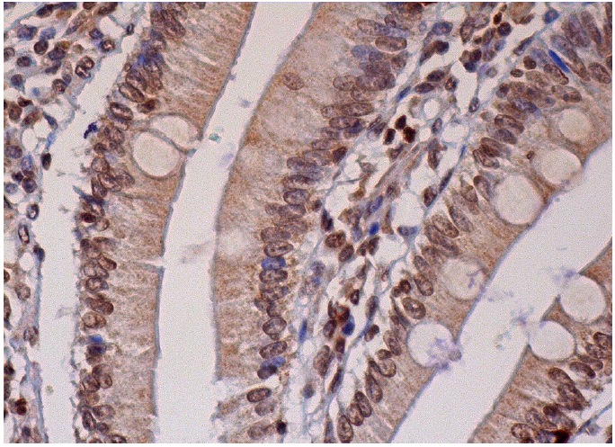
Immunoperoxidase staining of formalin fixed, paraffin-embedded human small intestine tissue showing cytoplasmic and nuclear staining of glandular cells.Application
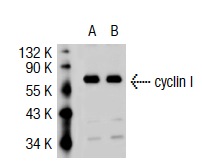
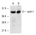
Western blot analysis of cyclin I expression in non-transfected: (A) and mouse cyclin I transfected: (B) 293T whole cell lysates.Application
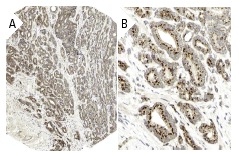
Immunoperoxidase staining of formalin fixed, paraffin-embedded human prostate cancer tissue showing cytoplasmic staining of tumor cells at low (A) and high (B) magnification. Kindly provided by The Swedish Human Protein Atlas (HPA) program.Application
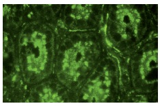
Immunofluor?escence staining of normal mouse intestine frozen section showing nuclear and cytoplasmic staining.Application
Western blot analysis of cyclin I expression in rat skeletal muscle (A) and mouse brain (B) extracts.| Product Name | cyclin I Antibody (H-279) |
|---|---|
| Antibody Type | Primary Antibodies |
| Antigen Alias | epitope corresponding to amino acids 1-279 mapping at the N-terminus of cyclin I of human origin |
| Modification Notes | Cyclins are the regulatory subunits of Cdc2 p34 and related cyclin-dependent kinases (Cdks) which play critical roles in the control of cell cycle progression. The catalytic subunit for cyclin A and B is Cdc2 p34 kinase. The Cdc2-cyclin B complex controls the G2 to M transition whereas Cdc2-cyclin A regulates S phase progression. Cyclin D1 accumulates during G1 and associates with Cdk2, Cdk4 and Cdk5. Cyclin E and Cdk2 interact during the G1 to S transition. Cyclin G contains a typical N-terminal cyclin box and a C-terminal domain sequence homologous to the tyrosine phosphorylation site of the epidermal growth factor receptor. Cyclin G2 shares 53% amino acid sequence identity with cyclin G1. Cyclin I shares highest sequence similarity to cyclins G and E and is most highly expressed in skeletal muscle, heart and brain. |
| Clonality | Polyclonal |
|---|---|
| Isotype | IgG |
| Host Species | Rabbit |
| Tested Applications | WBIPIFIHCELISA |
| recommended for detection of cyclin I of mouse, rat and human origin by WB, IP, IF, IHC(P) and ELISA also reactive with additional species, including equine, canine, bovine and porcine: |
|
| Species Reactivity | HumanMouseRat |
| Concentration | 1mg/ml |
| Purification | Affinity purified |
| Alternative Names | epitope corresponding to amino acids 1-279 mapping at the N-terminus of cyclin I of human origin |
|---|---|
| Tissue Specificity | epitope corresponding to amino acids 1-279 mapping at the N-terminus of cyclin I of human origin |
| Entrez Gene | 10983 |
|---|
Application
Immunoperoxidase staining of formalin fixed, paraffin-embedded human small intestine tissue showing cytoplasmic and nuclear staining of glandular cells.
Application
Western blot analysis of cyclin I expression in non-transfected: (A) and mouse cyclin I transfected: (B) 293T whole cell lysates.
Application
Immunoperoxidase staining of formalin fixed, paraffin-embedded human prostate cancer tissue showing cytoplasmic staining of tumor cells at low (A) and high (B) magnification. Kindly provided by The Swedish Human Protein Atlas (HPA) program.
Application
Immunofluor?escence staining of normal mouse intestine frozen section showing nuclear and cytoplasmic staining.
Application
Western blot analysis of cyclin I expression in rat skeletal muscle (A) and mouse brain (B) extracts.| Application Notes | recommended for detection of cyclin I of mouse, rat and human origin by WB, IP, IF, IHC(P) and ELISA also reactive with additional species, including equine, canine, bovine and porcine: |
|---|
| Form | Liquid |
|---|---|
| Storage Instructions | For short-term storage, store at 4° C. For long-term storage, aliquot and store at -20ºC or below. Avoid multiple freeze-thaw cycles. |
| Storage Buffer | phosphate buffered saline , pH 7.4, 150mM NaCl, 0.02% sodium azide and 50% glycerol. |


