Application
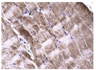
Immunoperoxidase staining of formalin fixed, paraffin-embedded human skeletal muscle tissue showing cytoplasmic staining of myocyte cells.Application
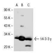
Western blot analysis of 14-3-3 γ expression in non-transfected 293T: (A), human 14-3-3 γ transfected 293T: (B) and A-431 (C) whole cell lysates.Application
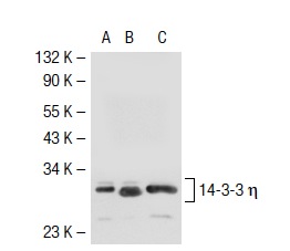
Western blot analysis of 14-3-3 η expression in non-transfected 293T: (A), mouse 14-3-3 η transfected 293T: (B) and NIH/3T3 (C) whole cell lysates.Application
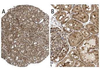
Immunoperoxidase staining of formalin fixed, paraffin-embedded human kidney tissue showing nuclear and cytoplasmic staining of cells in glomeruli and tubules (low and high magnification). Kindly provided by The Swedish Human Protein Atlas (HPA) program.Application
Western blot analysis of 14-3-3 γ expression in non-transfected 293T: (A), human 14-3-3 γ transfected 293T: (B) and K-562 (C) whole cell lysates.Application
Immunofluorescence staining of methanol-fixed A-431 cells showing cytoplasmic localization.Application
Western blot analysis of 14-3-3 γ expression in A-431 (A), K-562 (B), U-937 (C) and NIH/3T3 (D) whole cell lysates.| Product Name | 14-3-3 γ Antibody (C-16) |
|---|---|
| Antibody Type | Primary Antibodies |
| Modification Notes | 14-3-3 proteins regulate many cellular processes relevant to cancer biology, notably apoptosis, mitogenic signaling and cell-cycle checkpoints. Seven isoforms comprise this family of signaling intermediates, denoted 14-3-3 b, g, e, z, h, q and s. 14-3-3 proteins form dimers that present two binding sites for ligand proteins, thereby bringing together two proteins that may not otherwise associate. These ligands largely share a 14-3-3 consensus binding motif and exhibit serine/threonine phosphorylation. 14-3-3 proteins function in broad regulation of these ligand proteins; by cytoplasmic sequestration, occupation of interaction domains and import/export sequences, prevention of degradation, activation/repression of enzymatic activity, and facilitation of protein modification. Loss of expression contributes to a vast array of pathogenic cellular activities. |
| Clonality | Polyclonal |
|---|---|
| Isotype | IgG |
| Host Species | Rabbit |
| Tested Applications | WBIPIFIHCELISA |
| recommended for detection of 14-3-3 &gamma and, to a lesser extent, 14-3-3 &eta of mouse, rat and human origin by WB, IP, IF, IHC(P) and ELISA also reactive with additional species, including equine, canine, bovine, porcine and avian | |
| Species Reactivity | HumanMouseRat |
| Concentration | 1mg/ml |
| Purification | Affinity purified |
| Alternative Names | epitope mapping at the C-terminus of 14-3-3 &gamma of human origin |
|---|---|
| Tissue Specificity | epitope mapping at the C-terminus of 14-3-3 γ of human origin |
| Entrez Gene | 7532 |
|---|
Application
Immunoperoxidase staining of formalin fixed, paraffin-embedded human skeletal muscle tissue showing cytoplasmic staining of myocyte cells.
Application
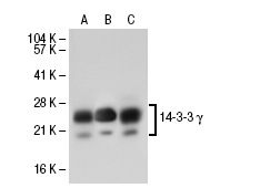
Western blot analysis of 14-3-3 γ expression in non-transfected 293T: (A), human 14-3-3 γ transfected 293T: (B) and A-431 (C) whole cell lysates.
Application
Western blot analysis of 14-3-3 η expression in non-transfected 293T: (A), mouse 14-3-3 η transfected 293T: (B) and NIH/3T3 (C) whole cell lysates.
Application
Immunoperoxidase staining of formalin fixed, paraffin-embedded human kidney tissue showing nuclear and cytoplasmic staining of cells in glomeruli and tubules (low and high magnification). Kindly provided by The Swedish Human Protein Atlas (HPA) program.
Application
Western blot analysis of 14-3-3 γ expression in non-transfected 293T: (A), human 14-3-3 γ transfected 293T: (B) and K-562 (C) whole cell lysates.
Application
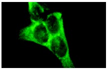
Immunofluorescence staining of methanol-fixed A-431 cells showing cytoplasmic localization.
Application
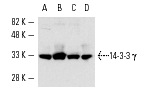
Western blot analysis of 14-3-3 γ expression in A-431 (A), K-562 (B), U-937 (C) and NIH/3T3 (D) whole cell lysates.| Application Notes | recommended for detection of 14-3-3 &gamma and, to a lesser extent, 14-3-3 &eta of mouse, rat and human origin by WB, IP, IF, IHC(P) and ELISA also reactive with additional species, including equine, canine, bovine, porcine and avian |
|---|
| Form | Liquid |
|---|---|
| Storage Instructions | For short-term storage, store at 4° C. For long-term storage, aliquot and store at -20ºC or below. Avoid multiple freeze-thaw cycles. |
| Storage Buffer | phosphate buffered saline , pH 7.4, 150mM NaCl, 0.02% sodium azide and 50% glycerol. |


