Application
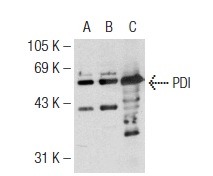
Western blot analysis of PDI expression in non-transfected 293: (A), human PDI transfected 293: (B) and Hep G2 (C) whole cell lysates.Application
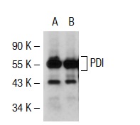
Western blot analysis of PDI expression in non-transfected: (A) and mouse PDI transfected: (B) 293T whole cell lysates.Application
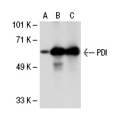
Western blot analysis of PDI expression in non-transfected 293T: (A), human PDI transfected 293T: (B) and Hep G2 (C) whole cell lysates.Application
Western blot analysis of PDI expression in non-transfected 293T: (A), human PDI transfected 293T: (B) and Hep G2 (C) whole cell lysatesApplication
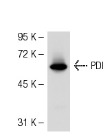
Western blot analysis of PDI expression in A-431 whole cell lysate.Application
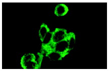
Immunofluorescence staining of methanol-fixed Hep G2 cells showing cytoplasmic localization.Application
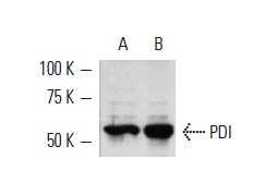
Western blot analysis of PDI expression in COLO 320DM (A) and Hep G2 (B) whole cell lysates.| Product Name | PDI Antibody (H-160) |
|---|---|
| Antibody Type | Primary Antibodies |
| Antigen Alias | epitope corresponding to amino acids 211-370 mapping to an internal region of PDI (protein disulfide isomerase) of human origin |
| Modification Notes | Oxidoreductase-protein disulfide isomerase (PDI) is a homodimer consisting of subunits that catalyzes thiol-disulfide exchange, mediates folding of newly synthesized proteins and functions as a molecular chaperone. PDI localizes to the lumen of the endoplasmic reticulum (ER), where in conjunction with folding-helper proteins, such as immunoglobulin heavy chain binding protein (BiP), mediates tertiary and quaternary protein-processing. Cell surface PDI induces sulfhydryl-mediated conformational changes in integrin-mediated adhesion receptor-ligand interactions, thereby regulating integrin responses and cell adhesion. Additionally, PDI functions as a subunit of two more complex enzyme systems: the prolyl-4-hydroxylase and the triacylglycerol transfer proteins. |
| Clonality | Polyclonal |
|---|---|
| Isotype | IgG |
| Host Species | Rabbit |
| Tested Applications | WBIPIFELISA |
| recommended for detection of PDI of mouse, rat and human origin by WB, IP, IF and ELISA also reactive with additional species, including equine, canine, bovine and avian: |
|
| Species Reactivity | HumanMouseRat |
| Concentration | 1mg/ml |
| Purification | Affinity purified |
| Alternative Names | epitope corresponding to amino acids 211-370 mapping to an internal region of PDI (protein disulfide isomerase) of human origin |
|---|---|
| Tissue Specificity | epitope corresponding to amino acids 211-370 mapping to an internal region of PDI (protein disulfide isomerase) of human origin |
| Entrez Gene | 5034 |
|---|
Application
Western blot analysis of PDI expression in non-transfected 293: (A), human PDI transfected 293: (B) and Hep G2 (C) whole cell lysates.
Application
Western blot analysis of PDI expression in non-transfected: (A) and mouse PDI transfected: (B) 293T whole cell lysates.
Application
Western blot analysis of PDI expression in non-transfected 293T: (A), human PDI transfected 293T: (B) and Hep G2 (C) whole cell lysates.
Application
Western blot analysis of PDI expression in non-transfected 293T: (A), human PDI transfected 293T: (B) and Hep G2 (C) whole cell lysates
Application
Western blot analysis of PDI expression in A-431 whole cell lysate.
Application
Immunofluorescence staining of methanol-fixed Hep G2 cells showing cytoplasmic localization.
Application
Western blot analysis of PDI expression in COLO 320DM (A) and Hep G2 (B) whole cell lysates.| Application Notes | recommended for detection of PDI of mouse, rat and human origin by WB, IP, IF and ELISA also reactive with additional species, including equine, canine, bovine and avian: |
|---|
| Form | Liquid |
|---|---|
| Storage Instructions | For short-term storage, store at 4° C. For long-term storage, aliquot and store at -20ºC or below. Avoid multiple freeze-thaw cycles. |
| Storage Buffer | phosphate buffered saline , pH 7.4, 150mM NaCl, 0.02% sodium azide and 50% glycerol. |


