Application
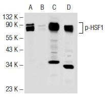
Antibodies tested include p-HSF1 (Ser 303): (A, B) and HSF1 (H-311): (C, D).Application
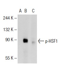
Western blot analysis of HSF1 phosphorylation in non-transfected: (A), untreated human HSF1 transfected: (B) and lambda protein phosphatase treated human HSF1 transfected: (C) 293T whole cell lysates.Application
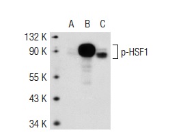
Western blot analysis of HSF1 phosphorylation in non-transfected 293T: (A), human HSF1 transfected 293T: (B) and HeLa (C) whole cell lysates.Application
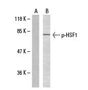
Blots were probed with p-HSF1 (Ser 303): preincubated with its cognate phosphorylated peptide (A) and p-HSF1 (Ser 303): (B).Application
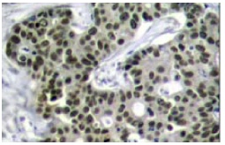
Immunoperoxidase staining of formalin-fixed, paraffin-embedded human breast carcinoma tissue showing nuclear localization.| Product Name | p-HSF1 Antibody (Ser 303) |
|---|---|
| Antibody Type | Primary Antibodies |
| Antigen Alias | epitope corresponding to phosphorylated Ser 303 of HSF1 of human origin |
| Modification | Phosphoralated |
| Modification Notes | Prokaryotic and eukaryotic cells respond to thermal and chemical stress by inducing a group of genes collectively designated heat shock genes. In eukaryotes, this gene expression is regulated primarily at the transcription level. Heat shock transcription factors (HSF, also designated HSTF) 1 and 2 are involved in this regulation. HSF1 and HSF2 are upregulated by estrogen, at both the mRNA and protein level. HSF1 is normally found as a monomer, whose transcriptional activity is repressed by constitutive phosphorylation. Upon activation, HSF1 forms trimers, gains DNA binding activity and is translocated to the nucleus. HSF2 activity is associated with differentiation and development, and, like HSF1, binds DNA as a trimer. Both HSF1 and HSF2 are known to be induced by proteasome inhibitors of the ubiquitin pathway. Phosphorylation of HSF1 on Ser230 by heat shock has been shown to positively contribute to the transcriptional activity of HSF1. The other phosphorylated serine residues, Ser 303, Ser 307 and Ser 363 have demonstrated repression of transactivation capacity. |
| Clonality | Polyclonal |
|---|---|
| Isotype | IgG |
| Host Species | Rabbit |
| Tested Applications | WBIPIFIHC |
| recommended for detection of Ser 303 phosphorylated HSF1 of h origin by WB, IP, IF and IHC(P): |
|
| Species Reactivity | HumanMouseRat |
| Concentration | 1mg/ml |
| Purification | Affinity purified |
| Alternative Names | epitope corresponding to phosphorylated Ser 303 of HSF1 of human origin |
|---|---|
| Tissue Specificity | epitope corresponding to phosphorylated Ser 303 of HSF1 of human origin |
| Entrez Gene | 3297 |
|---|
Application
Antibodies tested include p-HSF1 (Ser 303): (A, B) and HSF1 (H-311): (C, D).
Application
Western blot analysis of HSF1 phosphorylation in non-transfected: (A), untreated human HSF1 transfected: (B) and lambda protein phosphatase treated human HSF1 transfected: (C) 293T whole cell lysates.
Application
Western blot analysis of HSF1 phosphorylation in non-transfected 293T: (A), human HSF1 transfected 293T: (B) and HeLa (C) whole cell lysates.
Application
Blots were probed with p-HSF1 (Ser 303): preincubated with its cognate phosphorylated peptide (A) and p-HSF1 (Ser 303): (B).
Application
Immunoperoxidase staining of formalin-fixed, paraffin-embedded human breast carcinoma tissue showing nuclear localization.| Application Notes | recommended for detection of Ser 303 phosphorylated HSF1 of h origin by WB, IP, IF and IHC(P): |
|---|
| Form | Liquid |
|---|---|
| Storage Instructions | For short-term storage, store at 4° C. For long-term storage, aliquot and store at -20ºC or below. Avoid multiple freeze-thaw cycles. |
| Storage Buffer | phosphate buffered saline , pH 7.4, 150mM NaCl, 0.02% sodium azide and 50% glycerol. |


