Application
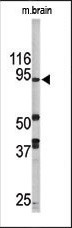
Western blot analysis of anti-LSD1 Pab (Cat. #AP1218a) in mouse brain tissue lysate (35ug/lane). LSD1(arrow) was detected using the purified Pab.Application
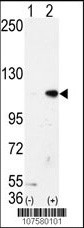
Western blot analysis of AOF2 (arrow) using LSD1 Antibody (N-term) (Cat.#AP1218a).293 cell lysates (2 ug/lane) either nontransfected (Lane 1) or transiently transfected with the AOF2 gene (Lane 2) (Origene Technologies).Application
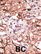
Formalin-fixed and paraffin-embedded human cancer tissue reacted with the primary antibody, which was peroxidase-conjugated to the secondary antibody, followed by DAB staining. This data demonstrates the use of this antibody for immunohistochemistry; clinical relevance has not been evaluated. BC = breast carcinoma; HC = hepatocarcinoma.Application
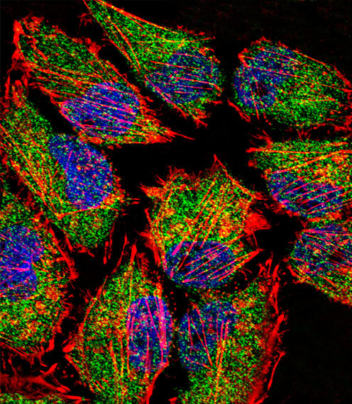
Fluorescent confocal image of Hela cell stained with LSD1 Antibody (N-term)(Cat#AP1218a).Hela cells were fixed with 4% PFA (20 min), permeabilized with Triton X-100 (0.1%, 10 min), then incubated with LSD1 primary antibody (1:25, 1 h at 37℃). For secondary antibody, Alexa Fluor® 488 conjugated donkey anti-rabbit antibody (green) was used (1:400, 50 min at 37℃).Cytoplasmic actin was counterstained with Alexa Fluor® 555 (red) conjugated Phalloidin (7units/ml, 1 h at 37℃). Nuclei were counterstained with DAPI (blue) (10 µg/ml, 10 min). LSD1 immunoreactivity is localized to Cytoplasm significantly and Nucleus weakly.| Product Name | LSD1 Antibody (N-term) |
|---|---|
| Antibody Type | Primary Antibodies |
| Antigen Alias | KDM1A; AOF2; KDM1; KIAA0601; LSD1; Lysine-specific histone demethylase 1A; BRAF35-HDAC complex protein BHC110; Flavin-containing amine oxidase domain-containing protein 2 |
| Clonality | Polyclonal |
|---|---|
| Isotype | Ig |
| Host Species | Rabbit |
| Tested Applications | WBIHCIF |
| WB:1:100~500 IHC |
|
| Species Reactivity | Human |
| Concentration | 1mg/ml |
| Gene Synonyms | AOF2 KDM1 KIAA0601 LSD1 |
|---|---|
| Alternative Names | KDM1A AOF2 KDM1 KIAA0601 LSD1 Lysine-specific histone demethylase 1A BRAF35-HDAC complex protein BHC110 Flavin-containing amine oxidase domain-containing protein 2 |
| Molecular Weight(MW) | 92903 Da |
| Function | Histone demethylase that demethylates both 'Lys-4' (H3K4me) and 'Lys-9' (H3K9me) of histone H3, thereby acting as a coactivator or a corepressor, depending on the context. Acts by oxidizing the substrate by FAD to generate the corresponding imine that is subsequently hydrolyzed. Acts as a corepressor by mediating demethylation of H3K4me, a specific tag for epigenetic transcriptional activation. Demethylates both mono- (H3K4me1) and di-methylated (H3K4me2) H3K4me. May play a role in the repression of neuronal genes. Alone, it is unable to demethylate H3K4me on nucleosomes and requires the presence of RCOR1/CoREST to achieve such activity. Also acts as a coactivator of androgen receptor (ANDR)-dependent transcription, by being recruited to ANDR target genes and mediating demethylation of H3K9me, a specific tag for epigenetic transcriptional repression. The presence of PRKCB in ANDR-containing complexes, which mediates phosphorylation of 'Thr- 6' of histone H3 (H3T6ph), a specific tag that prevents demethylation H3K4me, prevents H3K4me demethylase activity of KDM1A. Demethylates di-methylated 'Lys-370' of p53/TP53 which prevents interaction of p53/TP53 with TP53BP1 and represses p53/TP53-mediated transcriptional activation. Demethylates and stabilizes the DNA methylase DNMT1. Required for gastrulation during embryogenesis. Component of a RCOR/GFI/KDM1A/HDAC complex that suppresses, via histone deacetylase (HDAC) recruitment, a number of genes implicated in multilineage blood cell development Effector of SNAI1-mediated transcription repression of E- cadherin/CDH1, CDN7 and KRT8. Required for the maintenance of the silenced state of the SNAI1 target genes E-cadherin/CDH1 and CDN7 |
| Tissue Specificity | Ubiquitously expressed. |
| Cellular Localization | Nucleus. |
| Entrez Gene | 23028 |
|---|
Application
Western blot analysis of anti-LSD1 Pab (Cat. #AP1218a) in mouse brain tissue lysate (35ug/lane). LSD1(arrow) was detected using the purified Pab.
Application
Western blot analysis of AOF2 (arrow) using LSD1 Antibody (N-term) (Cat.#AP1218a).293 cell lysates (2 ug/lane) either nontransfected (Lane 1) or transiently transfected with the AOF2 gene (Lane 2) (Origene Technologies).
Application
Formalin-fixed and paraffin-embedded human cancer tissue reacted with the primary antibody, which was peroxidase-conjugated to the secondary antibody, followed by DAB staining. This data demonstrates the use of this antibody for immunohistochemistry; clinical relevance has not been evaluated. BC = breast carcinoma; HC = hepatocarcinoma.
Application
Fluorescent confocal image of Hela cell stained with LSD1 Antibody (N-term)(Cat#AP1218a).Hela cells were fixed with 4% PFA (20 min), permeabilized with Triton X-100 (0.1%, 10 min), then incubated with LSD1 primary antibody (1:25, 1 h at 37℃). For secondary antibody, Alexa Fluor® 488 conjugated donkey anti-rabbit antibody (green) was used (1:400, 50 min at 37℃).Cytoplasmic actin was counterstained with Alexa Fluor® 555 (red) conjugated Phalloidin (7units/ml, 1 h at 37℃). Nuclei were counterstained with DAPI (blue) (10 µg/ml, 10 min). LSD1 immunoreactivity is localized to Cytoplasm significantly and Nucleus weakly.| Application Notes | WB:1:100~500 IHC |
|---|
| Form | Liquid |
|---|---|
| Storage Instructions | For short-term storage, store at 4° C. For long-term storage, aliquot and store at -20ºC or below. Avoid multiple freeze-thaw cycles. |
| Storage Buffer | Purified polyclonal antibody supplied in PBS with 0.09% (W/V) sodium azide. This antibody is prepared by Saturated Ammonium Sulfate (SAS) precipitation followed by dialysis against PBS. |


