Application
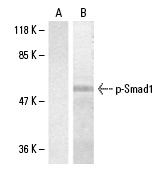
Western blot analysis of phosphorylated Smad1 expression in untreated (A) and PMA-treated (B) 293 whole cell lysates.| Product Name | p-Smad1 Antibody (Ser 465) |
|---|---|
| Antibody Type | Primary Antibodies |
| Antigen Alias | epitope corresponding to phosphorylated Ser 465 of Smad1 of human origin |
| Modification | Phosphoralated |
| Modification Notes | Smad proteins, the mammalian homologs of the Drosophila Mothers against dpp (Mad), function downstream of TGF∫ receptor serine/threonine kinases and undergo serine phosphorylation in response to receptor activation (1,2). Following BMP (bone morphogenic protein) or TGF∫ binding to the targeted surface receptors, Smad1 (also designated Madr1 or JV4-1) becomes phosphorylated at Ser-463 and Ser-465 (3–5). Activated Smad1 associates with Smad4 and translocates to the nucleus, where the Smad1-Smad4 complex recruits DNA-binding proteins to activate specific gene transcription (3,4). BMP binding induces phosphorylation of Smad1, which enhances the binding of Smad1 to CBP to stimulate Smad1-dependent transcription (1,2). In addition, ALK1 and ALK2 specifically phosphorylate Smad1 on residues located in the MH2 domain and induces Smad1-dependent pathways (6,7). |
| Clonality | Polyclonal |
|---|---|
| Isotype | IgG |
| Host Species | Rabbit |
| Tested Applications | WBIP |
| recommended for detection of Ser 465 phosphorylated Smad1 of human and mouse origin and correspondingly phosphorylated Ser 468 of rat origin by WB and IP: |
|
| Species Reactivity | HumanMouseRat |
| Concentration | 1mg/ml |
| Purification | Affinity purified |
| Alternative Names | epitope corresponding to phosphorylated Ser 465 of Smad1 of human origin |
|---|---|
| Tissue Specificity | epitope corresponding to phosphorylated Ser 465 of Smad1 of human origin |
| Entrez Gene | 4086 |
|---|
Application
Western blot analysis of phosphorylated Smad1 expression in untreated (A) and PMA-treated (B) 293 whole cell lysates.| Application Notes | recommended for detection of Ser 465 phosphorylated Smad1 of human and mouse origin and correspondingly phosphorylated Ser 468 of rat origin by WB and IP: |
|---|
| Form | Liquid |
|---|---|
| Storage Instructions | For short-term storage, store at 4° C. For long-term storage, aliquot and store at -20ºC or below. Avoid multiple freeze-thaw cycles. |
| Storage Buffer | phosphate buffered saline , pH 7.4, 150mM NaCl, 0.02% sodium azide and 50% glycerol. |


