Application
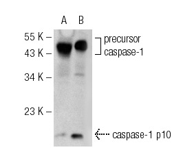
Western blot analysis of group caspase-1 p10 expression in RAW 309 Cr.1 (A) and RAW 309 Cr.1 & LPS (B) whole cell lysates.Application
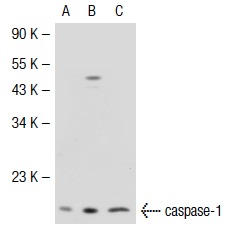
Western blot analysis of caspase-1 expression in non-transfected 293T: (A), mouse caspase-1 transfected 293T: (B) and WEHI-231 (C) whole cell lysates.Application
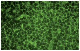
Immunofluorescence staining of normal mouse lymph node frozen section showing cytoplasmic staining.Application
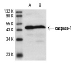
Western blot analysis of caspase-1 precursor expression in LPS-induced RAW 309 whole cell lysate (A) and mouse spleen extract (B).Application
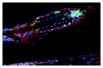
Cryostat section of mouse skin showing a single hair follicle staining with caspase-1 p10 (red immunofluorescence staining). Note also TUNEL staining marking apoptotic cells (green fluorescence) and HOECHST 33342 nuclear counterstain (blue fluorescence). Kindly provided by Hair Research Group, Humboldt University, Berlin.WB
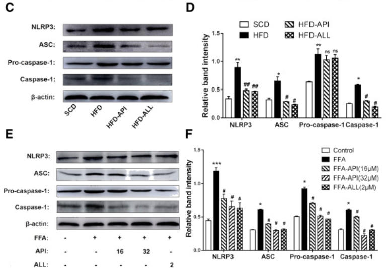
(C-D) NLRP3, ASC, pro-caspase-1, caspase-1 protein levels were detected by Western blots and quantified by using Image J software in mice. The data are expressed as the mean±S.E.M. (n=6). *P<.05, **P<.01, ***P<.01 vs. The SCD group; #P<.05 ##P<.01 vs. the HFD group. (E-F) NLRP3, ASC, Pro-caspase-1, caspase-1 protein levels were detected by Western blots and quantified by using Image J software in hepa1–6 cells.| Product Name | caspase-1 p10 Antibody (M-20) |
|---|---|
| Antibody Type | Primary Antibodies |
| Product description |
|
| Modification Notes | Caspase-1, originally designated ICE (for IL-1 converting enzyme), is a member of the group of caspases with large prodomains. Caspase-1 promotes maturation of interleukin IL-1 beta and interleukin18 (IL-18) by proteolytic cleavage of precursor forms into biologically active pro-inflamatory cytokines. The prodomain of caspase-1 (also known as Pro-C1) represents the amino acid terminal portion of the caspase-1 precursor. Pro-C1 is produced as a residual component after proteolytic cleavage of the precursor generates the functional caspase-1 subunits known as the p20 and p10 subunits. Active caspase-1, a (p20/p10)2 tetramer, is necessary and sufficient for cleavage of precursor IL-1 as well as for induction of apoptosis in some cell lines. The highly conserved family of caspases mediate many of the morphological and biochemical features of apoptosis, including structural dismantling of cell bodies and nuclei, fragmentation of genomic DNA, destruction of regulatory proteins and propagation of other pro-apoptotic molecules. The human Caspase-1 gene maps to chromosome 2q14 and encodes a cytoplasmic protein expressed in liver, heart, skeletal muscle kidney and testis. Caspase-1 has been implicated in inflammation, septic shock, and other situations such as wound healing and the growth of certain leukemias. |
| Clonality | Polyclonal |
|---|---|
| Isotype | IgG |
| Host Species | Rabbit |
| Tested Applications | ELISAIFIPWB |
| recommended for detection of the p10 subunit and precursor of caspase-1 of mouse and rat origin by WB, IP, IF and ELISA non cross-reactive with caspase-1 p20 also reactive with additional species, including equine and porcine: |
|
| Species Reactivity | HumanMouseRat |
| Concentration | 1mg/ml |
| Purification | Affinity purified |
| Alternative Names | epitope mapping at the C-terminus of caspase-1 p10 of mouse origin |
|---|---|
| Tissue Specificity | epitope mapping at the C-terminus of caspase-1 p10 of mouse origin |
| Entrez Gene | 12362 |
|---|
Application
Western blot analysis of group caspase-1 p10 expression in RAW 309 Cr.1 (A) and RAW 309 Cr.1 & LPS (B) whole cell lysates.
Application
Western blot analysis of caspase-1 expression in non-transfected 293T: (A), mouse caspase-1 transfected 293T: (B) and WEHI-231 (C) whole cell lysates.
Application
Immunofluorescence staining of normal mouse lymph node frozen section showing cytoplasmic staining.
Application
Western blot analysis of caspase-1 precursor expression in LPS-induced RAW 309 whole cell lysate (A) and mouse spleen extract (B).
Application
Cryostat section of mouse skin showing a single hair follicle staining with caspase-1 p10 (red immunofluorescence staining). Note also TUNEL staining marking apoptotic cells (green fluorescence) and HOECHST 33342 nuclear counterstain (blue fluorescence). Kindly provided by Hair Research Group, Humboldt University, Berlin.
WB
(C-D) NLRP3, ASC, pro-caspase-1, caspase-1 protein levels were detected by Western blots and quantified by using Image J software in mice. The data are expressed as the mean±S.E.M. (n=6). *P<.05, **P<.01, ***P<.01 vs. The SCD group; #P<.05 ##P<.01 vs. the HFD group. (E-F) NLRP3, ASC, Pro-caspase-1, caspase-1 protein levels were detected by Western blots and quantified by using Image J software in hepa1–6 cells.| Application Notes | recommended for detection of the p10 subunit and precursor of caspase-1 of mouse and rat origin by WB, IP, IF and ELISA non cross-reactive with caspase-1 p20 also reactive with additional species, including equine and porcine: |
|---|
| Form | Liquid |
|---|---|
| Storage Instructions | For short-term storage, store at 4° C. For long-term storage, aliquot and store at -20ºC or below. Avoid multiple freeze-thaw cycles. |
| Storage Buffer | phosphate buffered saline , pH 7.4, 150mM NaCl, 0.02% sodium azide and 50% glycerol. |


