Application
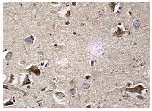
Immunoperoxidase staining of formalin fixed, paraffin-embedded human brain tissue showing cytoplasmic and nuclear staining of neuronal cells.Application
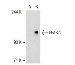
Western blot analysis of EPAS-1 expression in non-transfected: (A) and mouse EPAS-1 transfected: (B) 293T whole cell lysates.Application
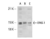
Western blot analysis of EPAS-1 expression in HT-1080 (A), CoCl2-induced HeLa (B) and A549 (C) whole cell lysates.| Product Name | EPAS-1 Antibody (H-310) |
|---|---|
| Antibody Type | Primary Antibodies |
| Antigen Alias | epitope corresponding to amino acids 556-865 mapping near the C-terminus of EPAS-1 of human origin |
| Modification Notes | Cell growth and viability is compromised by oxygen deprivation (hypoxia). Hypoxia-inducible factors, including HIF-1a, HIF-1b (also designated Arnt 1), EPAS-1 (also designated HIF-2a) and HIF-3a, induce glycolysis, erythropoiesis and angiogenesis in order to restore oxygen homeostasis. Hypoxia-inducible factors are members of the Per-Arnt-Sim (PAS) domain transcription factor family. In response to hypoxia, HIF-1a is upregulated and forms a heterodimer with Arnt 1 to form the HIF-1 complex. The HIF-1 complex recognizes and binds to the hypoxia responsive element (HRE) of hypoxia-inducible genes, thereby activating transcription. Hypoxia-inducible expression of some genes such as Glut-1, p53, p21 or Bcl-2, is HIF-1a dependent, whereas expression of others, such as p27, GADD 153 or HO-1, is HIF-1a independent. EPAS-1 and HIF-3a have also been shown to form heterodimeric complexes with Arnt 1 in response to hypoxia. |
| Clonality | Polyclonal |
|---|---|
| Isotype | IgG |
| Host Species | Rabbit |
| Tested Applications | WBIPIFIHCELISAGSChIP |
| recommended for detection of EPAS-1 of mouse, rat and human origin by WB, IP, IF, IHC(P) and ELISA: |
|
| Species Reactivity | HumanMouseRat |
| Concentration | 1mg/ml |
| Purification | Affinity purified |
| Alternative Names | epitope corresponding to amino acids 556-865 mapping near the C-terminus of EPAS-1 of human origin |
|---|---|
| Tissue Specificity | epitope corresponding to amino acids 556-865 mapping near the C-terminus of EPAS-1 of human origin |
| Entrez Gene | 2034 |
|---|
Application
Immunoperoxidase staining of formalin fixed, paraffin-embedded human brain tissue showing cytoplasmic and nuclear staining of neuronal cells.
Application
Western blot analysis of EPAS-1 expression in non-transfected: (A) and mouse EPAS-1 transfected: (B) 293T whole cell lysates.
Application
Western blot analysis of EPAS-1 expression in HT-1080 (A), CoCl2-induced HeLa (B) and A549 (C) whole cell lysates.| Application Notes | recommended for detection of EPAS-1 of mouse, rat and human origin by WB, IP, IF, IHC(P) and ELISA: |
|---|
| Form | Liquid |
|---|---|
| Storage Instructions | For short-term storage, store at 4° C. For long-term storage, aliquot and store at -20ºC or below. Avoid multiple freeze-thaw cycles. |
| Storage Buffer | phosphate buffered saline , pH 7.4, 150mM NaCl, 0.02% sodium azide and 50% glycerol. |


