Application
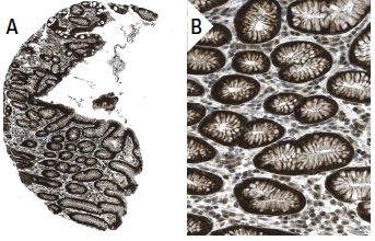
Immunoperoxidase staining of formalin fixed, paraffin-embedded human stomach tissue showing cytoplasmic staining of glandular cells (low and high magnification). Kindly provided by The Swedish Human Protein Atlas (HPA) program.Application
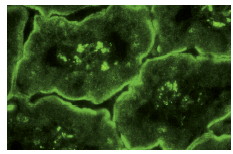
Immunofluorescence staining of normal mouse intestine frozen section showing membrane staining.| Product Name | DDEF2 Antibody (H-300) |
|---|---|
| Antibody Type | Primary Antibodies |
| Antigen Alias | AMAP2,ASAP2,PAG3,PAP,SHAG1 |
| Conjugation | Agarose |
| Modification | Acetylated |
| Modification Notes | DDEF2 (ADP ribosylation factor [ARF]- GTPase-activating protein [GAP] containing SH3, ANK repeats, and PH domain, PAP, PAG2, AMAP1, ZG14P, centaurin beta4) is a phospholipid-dependent ADP-ribosylation factor (ARF) GTPase-activating protein (ARF-GAP) that binds to protein-tyrosine kinases Src and focal adhesion kinase. ARF family GTP-binding proteins are regulators of membrane traffic and cytoskeletal organization. Modulation of ARF activity by DDEF2 is important for the regulation of focal adhesion assembly and/or organization by influencing the mechanisms responsible for the recruitment and organization of focal adhesion proteins paxillin and FAK. In spreading platelets, most endogenous DDEF2 is localized at peripheral focal adhesions. Pyk2 directly phosphorylates DDEF2 on tyrosine-308 and -782, and this event affects the phosphoinositide binding profile of DDEF2. DDEF2 is phosphorylated on tyrosine residues in cells expressing activated Src and tyrosine phosphorylation depends on a proline-rich class II Src SH3 binding site. |
| Clonality | Polyclonal |
|---|---|
| Isotype | IgG |
| Host Species | Rabbit |
| Tested Applications | ELISAIFIHCIPWB |
| WB:1:100~1:400 IP:1:20~1:100 IF:1:20~1:100 IHC:1:10~1:50 |
|
| Species Reactivity | HumanMouseRat |
| Concentration | 1mg/ml |
| Purification | Affinity purified |
| Alternative Names | AMAP2 ASAP2 PAG3 PAP SHAG1 |
|---|---|
| Molecular Weight(MW) | 112 kDa |
| Tissue Specificity | epitope corresponding to amino acids 1-300 mapping at the N-terminus of DDEF2 of human origin |
Application
Immunoperoxidase staining of formalin fixed, paraffin-embedded human stomach tissue showing cytoplasmic staining of glandular cells (low and high magnification). Kindly provided by The Swedish Human Protein Atlas (HPA) program.
Application
Immunofluorescence staining of normal mouse intestine frozen section showing membrane staining.| Application Notes | WB:1:100~1:400 IP:1:20~1:100 IF:1:20~1:100 IHC:1:10~1:50 |
|---|
| Form | Liquid |
|---|---|
| Storage Instructions | For short-term storage, store at 4° C. For long-term storage, aliquot and store at -20ºC or below. Avoid multiple freeze-thaw cycles. |
| Storage Buffer | phosphate buffered saline , pH 7.4, 150mM NaCl, 0.02% sodium azide and 50% glycerol. |


