Application
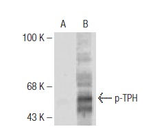
Western blot analysis of untreated (A) and cAMP-dependent protein kinase (PKA)-treated (B) rabbit recombinant TPH.| Product Name | p-TPH Antibody (Ser 58) |
|---|---|
| Antibody Type | Primary Antibodies |
| Modification Notes | Phenylalanine hydroxylase (PAH), tyrosine hydroxylase (TH) and tryptophan hydroxylase (TPH) comprise a small family of monooxygenases that use tetrahydropterine as a cofactor during the catabolism of aromatic L-amino acids (1,2). PAH, TH and TPH all contain catalytic domains with an amino-terminal regulatory domain and a short carboxy-terminal tetramerization domain (1). Each of these enzymes also contains a single ferrous iron atom, which is bound to two histidines and a glutamate and is likely to be involved in the formation of the hydroxylating intermediate (1). TPH is the first and rate-limiting step in the biosynthesis of serotonin in the central nervous system and melatonin in the pineal gland (1-5). Alteration of TPH function may be a key factor in the pathology of several neuropsychiatric disorders associated with serotonin, including depression, aggression, alcoholism and schizophrenia (3,4,6,7). For instance, L-DOPA, which is used as a common therapy for Parkinson’s disease (PD) patients, inhibits TPH function, which subsequently, is thought to contribute to the onset of depression in PD patients (3,4). Mouse, rat, human and rabbit TPH undergo PKA-mediated phosphorylation on Ser 58, a residue that is located in the N-terminal regulatory domain. |
| Clonality | Polyclonal |
|---|---|
| Isotype | IgG |
| Host Species | Rabbit |
| Tested Applications | WBIP |
| Species Reactivity | HumanMouseRat |
| Concentration | 1mg/ml |
| Purification | Affinity purified |
| Alternative Names | recommended for detection of Ser 58 phosphorylated TPH of rat human and rabbit origin and correspondingly Ser 61 phosphorylated TPH of mouse origin of mouse rat human rabbit Xenopus and zebrafish origin by WB and IP also reactive with additional spec |
|---|---|
| Tissue Specificity | recommended for detection of Ser 58 phosphorylated TPH of rat, human and rabbit origin and correspondingly Ser 61 phosphorylated TPH of mouse origin of mouse, rat, human, rabbit, Xenopus and zebrafish origin by WB and IP; also reactive with additional species, including bovine and canine |
| Entrez Gene | 7166 |
|---|
Application
Western blot analysis of untreated (A) and cAMP-dependent protein kinase (PKA)-treated (B) rabbit recombinant TPH.| Form | Liquid |
|---|---|
| Storage Instructions | For short-term storage, store at 4° C. For long-term storage, aliquot and store at -20ºC or below. Avoid multiple freeze-thaw cycles. |
| Storage Buffer | phosphate buffered saline , pH 7.4, 150mM NaCl, 0.02% sodium azide and 50% glycerol. |


