Application
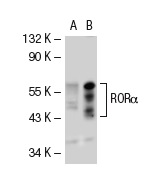
Western blot analysis of RORα expression in non-transfected: (A) and mouse RORα transfected: (B) 293T whole cell lysates.Application
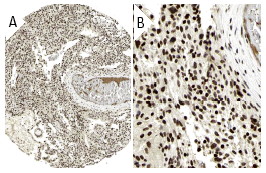
Immunoperoxidase staining of formalin fixed, paraffin-embedded human placenta tissue showing nuclear staining of decidual and trophoblastic cells at low (A) and high (B) magnification. Kindly provided by The Swedish Human Protein Atlas (HPA) program.Application
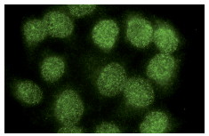
Immunofluorescence staining of methanol-fixed Hela cells showing nuclear localization.Application
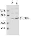
Western blot analysis of RORα expression in rat cerebellum (A) and mouse cerebellum (B) tissue extracts.Application
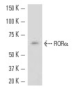
Western blot analysis of RORα expression in mouse cerebellum tissue extract.IHC
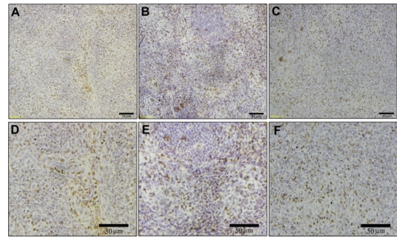
The immunostaining of the 3 melatonin nuclear receptors RORα (A, D), RORβ (B, E) and RORγ (C, F) in the spleen of broilers at 14 D.IHC
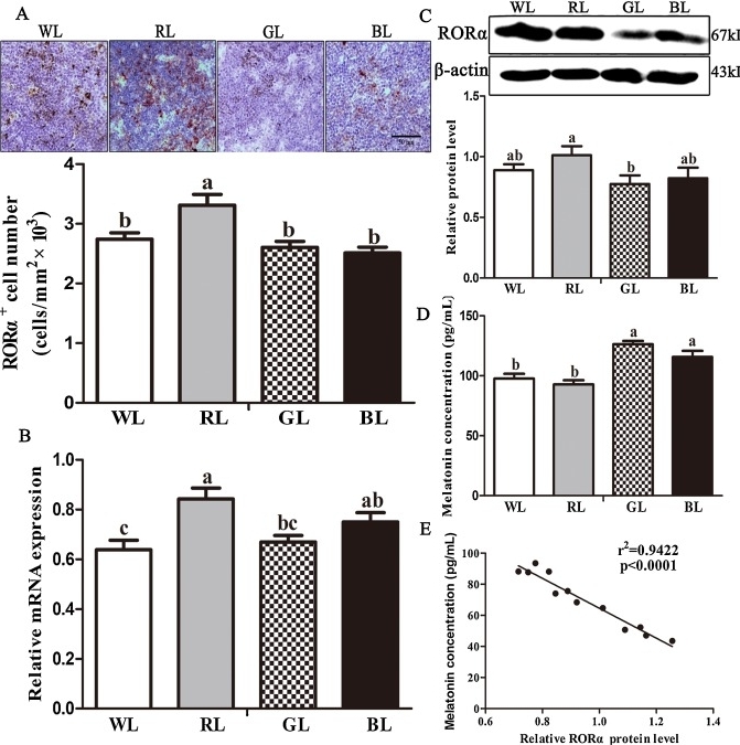
A: RORα-positive cells; B: RORα mRNA expression; C: RORα protein expression; D: plasma melatonin concentration; E: correlation between the melatonin level and RORα protein level.IHC
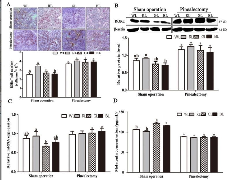
A: RORα-positive cells; B: RORα protein expression; C: RORα mRNA expression; D: plasma melatonin concentration.| Product Name | RORα Antibody (H-65) |
|---|---|
| Antibody Type | Primary Antibodies |
| Conjugation | Agarose |
| Modification | Acetylated |
| Modification Notes | Retinoids are metabolites of vitamin A (retinol) and represent an important class of signaling molecule during vertebrate development and tissue differentiation. A large group of nuclear transcription factors, including vitamin D3 receptor (VDR), thyroid hormone receptor (TR), RAR, RXR and ecdysone receptor, have a high affinity for retinoic acids and are members of the steroid receptor superfamily. Members of this family act by directly associating with DNA sequences known as hormone response elements (HREs) and bind DNA as either homo- or heterodimers. RORå is a member of the steroid receptor superfamily and is classified as an “orphan receptor” due to the lack of a defined ligand. Two isoforms of RORå have been described and are designated RORå1 and RORå2. RORå, also referred to as RZR, binds DNA as a monomer at consensus RORå response elements (ROREs). |
| Clonality | Polyclonal |
|---|---|
| Isotype | IgG |
| Host Species | Rabbit |
| Tested Applications | ChIPELISAGSIFIHCIPWB |
| WB:1:100~1:400 IP:1:20~1:100 IF:1:20~1:100 IHC:1:10~1:50 |
|
| Species Reactivity | HumanMouseRat |
| Concentration | 1mg/ml |
| Purification | Affinity purified |
| Alternative Names | epitope corresponding to amino acids 196-260 mapping within an internal region of RORα2 of human origin |
|---|---|
| Tissue Specificity | epitope corresponding to amino acids 196-260 mapping within an internal region of RORα2 of human origin |
| Entrez Gene | 6095 |
|---|
Application
Western blot analysis of RORα expression in non-transfected: (A) and mouse RORα transfected: (B) 293T whole cell lysates.
Application
Immunoperoxidase staining of formalin fixed, paraffin-embedded human placenta tissue showing nuclear staining of decidual and trophoblastic cells at low (A) and high (B) magnification. Kindly provided by The Swedish Human Protein Atlas (HPA) program.
Application
Immunofluorescence staining of methanol-fixed Hela cells showing nuclear localization.
Application
Western blot analysis of RORα expression in rat cerebellum (A) and mouse cerebellum (B) tissue extracts.
Application
Western blot analysis of RORα expression in mouse cerebellum tissue extract.
IHC
The immunostaining of the 3 melatonin nuclear receptors RORα (A, D), RORβ (B, E) and RORγ (C, F) in the spleen of broilers at 14 D.
IHC
A: RORα-positive cells; B: RORα mRNA expression; C: RORα protein expression; D: plasma melatonin concentration; E: correlation between the melatonin level and RORα protein level.
IHC
A: RORα-positive cells; B: RORα protein expression; C: RORα mRNA expression; D: plasma melatonin concentration.| Application Notes | WB:1:100~1:400 IP:1:20~1:100 IF:1:20~1:100 IHC:1:10~1:50 |
|---|
| Form | Liquid |
|---|---|
| Storage Instructions | For short-term storage, store at 4° C. For long-term storage, aliquot and store at -20ºC or below. Avoid multiple freeze-thaw cycles. |
| Storage Buffer | phosphate buffered saline , pH 7.4, 150mM NaCl, 0.02% sodium azide and 50% glycerol. |


