Application
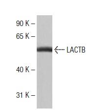
Western blot analysis of LACTB expression in human fetal liver tissue extract.Application
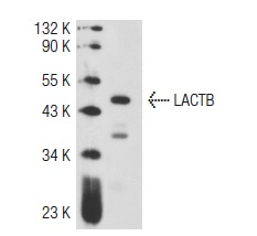
Western blot analysis of LACTB expression in HeLa whole cell lysate.IF
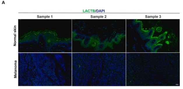
(A): LACTB expression in melanoma tissue chip and relative normal skin was measured by immunofluorescence staining.WB
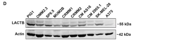
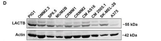
(D): The protein levels of LACTB were detected by western blotting in melanoma cell lines.WB
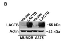
(B): Overexpression of LACTB protein in MUM2B and A375 cells was assessed by western blotting.IF
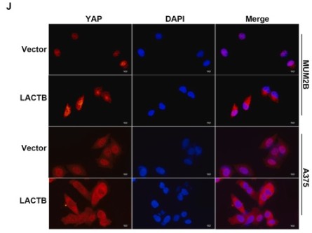
(J): Immunofluorescence staining of YAP revealed its cytoplasmic retention caused by ectopic LACTB expression in MUM2B and A375 cells. Left panel, representative images; Right panel, statistical graph. Scale bar, 100 μm.WB
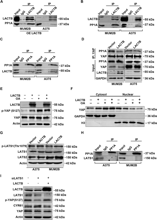
(A): A co-IP assay was carried out in MUM2B and A375 cells overexpressing LACTB using anti-LACTB. IgG was used as a negative control. (B): Endogenous LACTB interacted with PP1A in MUM2B and A375 cells. (C): A co-IP assay was carried out with an antibody against PP1A in MUM2B and A375 cells. (D): The indicated cells were immunoprecipitated using an antibody against YAP before detection of PP1A. The overexpression of LACTB suppressed the interaction of PP1A with YAP. (E): MUM2B cells overexpressing LACTB or the vector control were treated with 100 nM okadaic acid for 4 fours. Cell lysates were collected for detection of indicated proteins. (F): YAP subcellular localization was determined. (G): Western blotting analysis of p-LATS1 (Thr1079), LATS1 and LATS2 in the indicated cells. (H): The interaction between PP1A and LAST1 in A735 and MUM2B cells was investigated by co-IP. (I): MUM2B cells with or without stable LACTB overexpression were transfected with siNC or siLATS1. Then, cell lysates were harvested two days later and used for the detection of the indicated proteins.WB
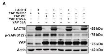
(A): MUM2B cells overexpressing LACTB or empty vector were transfected with the indicated plasmids and then subjected to SDS-PAGE for immunoblotting analysis of LACTB, p-YAP (S127) and YAP.WB
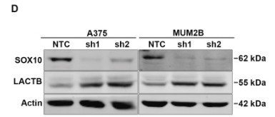
(D): Results of western blotting using primary antibodies against SOX10 and LACTB in the indicated cells.WB
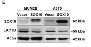
(E): The two cell lines were infected with lentiviral particles for the stable overexpression of SOX10. Then the levels of SOX10 and LACTB in the indicated cells were detected by western blotting.WB
(D): The protein levels of LACTB were detected by western blotting in melanoma cell lines.| Product Name | LACTB Antibody (O-21) |
|---|---|
| Antibody Type | Primary Antibodies |
| Modification Notes | Penicillin refers to any member of beta-lactam antibiotics group. These agents are identified by a beta-lactam ring within their molecular structure. As the most widely used group of antibiotics available, beta-lactams are used for the treatment of bacterial infections usually caused by gram-positive organisms. Beta-lactam antibiotics are bactericidal, functioning to inhibit the synthesis of the peptidoglycan layer of bacterial cell walls. Bacterial penicillin-binding proteins and beta-lactamases constitute a large family of serine proteases that perform essential functions in the synthesis and maintenance of peptidoglycan cell wall. Notably, beta-lactamases cleave beta-lactams, therefore providing the bacteria with resistance to the antibiotic. Homologues of beta-lactamases occur in many species, including human, rat, cow, rabbit, pig, xenopus, zebrafish, and C. elegans. The human homologues, LACTB and LACTB2, are active-site-serine enzymes thought to be involved in metabolism. |
| Clonality | Polyclonal |
|---|---|
| Isotype | IgG |
| Host Species | Rabbit |
| Tested Applications | ELISAIFIPWB |
| WB:1:500~1:2000 IP:1:100~1:500 Notes:Optimal dilutions/concentrations should be determined by the researcher. |
|
| Species Reactivity | HumanMouseRat |
| Concentration | 1mg/ml |
| Purification | Affinity purified |
| Alternative Names | recommended for detection of LACTB of mouse rat and human origin by WB IP and ELISA |
|---|---|
| Tissue Specificity | recommended for detection of LACTB of mouse, rat and human origin by WB, IP and ELISA |
| Entrez Gene | 114294 |
|---|
Application
Western blot analysis of LACTB expression in human fetal liver tissue extract.
Application
Western blot analysis of LACTB expression in HeLa whole cell lysate.
IF
(A): LACTB expression in melanoma tissue chip and relative normal skin was measured by immunofluorescence staining.
WB
(D): The protein levels of LACTB were detected by western blotting in melanoma cell lines.
WB
(B): Overexpression of LACTB protein in MUM2B and A375 cells was assessed by western blotting.
IF
(J): Immunofluorescence staining of YAP revealed its cytoplasmic retention caused by ectopic LACTB expression in MUM2B and A375 cells. Left panel, representative images; Right panel, statistical graph. Scale bar, 100 μm.
WB
(A): A co-IP assay was carried out in MUM2B and A375 cells overexpressing LACTB using anti-LACTB. IgG was used as a negative control. (B): Endogenous LACTB interacted with PP1A in MUM2B and A375 cells. (C): A co-IP assay was carried out with an antibody against PP1A in MUM2B and A375 cells. (D): The indicated cells were immunoprecipitated using an antibody against YAP before detection of PP1A. The overexpression of LACTB suppressed the interaction of PP1A with YAP. (E): MUM2B cells overexpressing LACTB or the vector control were treated with 100 nM okadaic acid for 4 fours. Cell lysates were collected for detection of indicated proteins. (F): YAP subcellular localization was determined. (G): Western blotting analysis of p-LATS1 (Thr1079), LATS1 and LATS2 in the indicated cells. (H): The interaction between PP1A and LAST1 in A735 and MUM2B cells was investigated by co-IP. (I): MUM2B cells with or without stable LACTB overexpression were transfected with siNC or siLATS1. Then, cell lysates were harvested two days later and used for the detection of the indicated proteins.
WB
(A): MUM2B cells overexpressing LACTB or empty vector were transfected with the indicated plasmids and then subjected to SDS-PAGE for immunoblotting analysis of LACTB, p-YAP (S127) and YAP.
WB
(D): Results of western blotting using primary antibodies against SOX10 and LACTB in the indicated cells.
WB
(E): The two cell lines were infected with lentiviral particles for the stable overexpression of SOX10. Then the levels of SOX10 and LACTB in the indicated cells were detected by western blotting.
WB
(D): The protein levels of LACTB were detected by western blotting in melanoma cell lines.| Application Notes | WB:1:500~1:2000 IP:1:100~1:500 Notes:Optimal dilutions/concentrations should be determined by the researcher. |
|---|
| Form | Liquid |
|---|---|
| Storage Instructions | For short-term storage, store at 4° C. For long-term storage, aliquot and store at -20ºC or below. Avoid multiple freeze-thaw cycles. |
| Storage Buffer | phosphate buffered saline , pH 7.4, 150mM NaCl, 0.02% sodium azide and 50% glycerol. |


