


Application
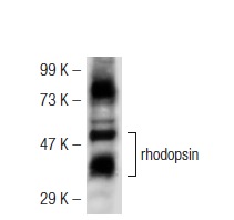
Western blot analysis of rhodopsin expression in mouse eye tissue extract.IF
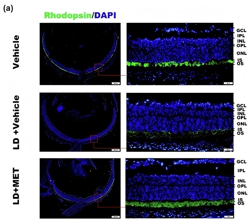
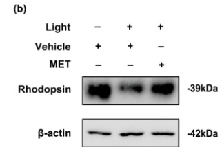
Administration of metformin maintained the protein level of rhodopsin. The mice were intraperitoneally injected with metformin and exposed to the light conditions for 12 h. (a). The retinal frozen sections were immunostained for detecting the level of rhodopsin. Scale bar = 200 μm; 50 μm.WB
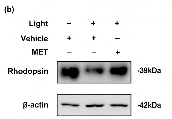
(b). The retinas were harvested and lysed for the western blot assay and β-actin was used as an internal control.WB
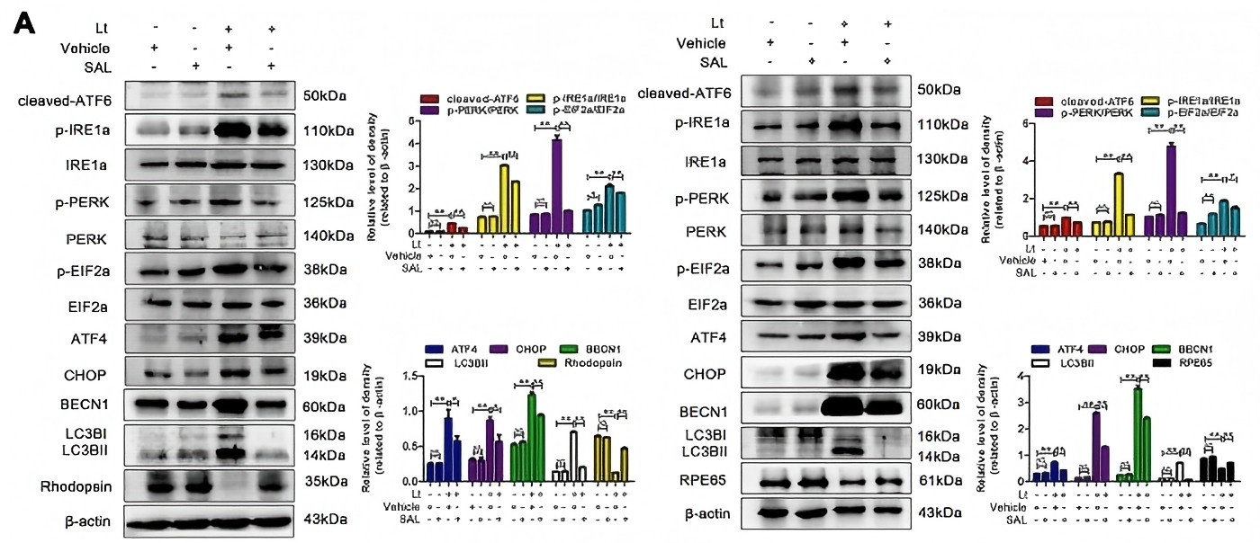
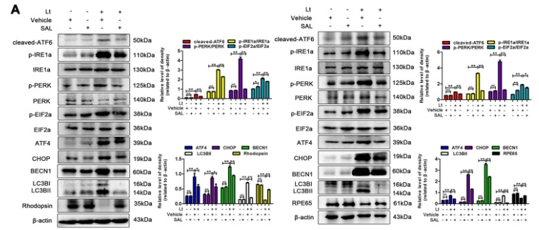
(A) The retinas were collected, and target proteins were determined with western blotting. β-actin was referenced as an internal control. Three independent experiments are conducted three weeks apart. The results are presented as the mean± SEM. n (per group) =3, NS: no significance, *P < 0.05, **P < 0.01.IF
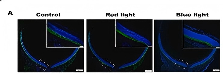
A. Immunofluorescence of rhodopsin (blue: DAPI, green: Rhodopsin, scale bar = 50 μm).WB
(A) The retinas were collected, and target proteins were determined with western blotting. β-actin was referenced as an internal control. Three independent experiments are conducted three weeks apart. The results are presented as the mean± SEM. n (per group) =3, NS: no significance, *P < 0.05, **P < 0.01.IF
(a). The retinal frozen sections were immunostained for detecting the level of rhodopsin. Scale bar = 200 μm; 50 μm.WB
(b). The retinas were harvested and lysed for the western blot assay and β-actin was used as an internal control.IF
(c). The immunofluorescence intensity of rhodopsin was then quantitatively analyzed. (d). The protein level of rhodopsin was quantitatively analyzed. Each experiment was repeated at least 3 times. The results have been presented as means ± SEM, *p < 0.05.IF
A. Immunofluorescence of rhodopsin (blue: DAPI, green: Rhodopsin, scale bar = 50 μm).IF
A. Immunofluorescence of rhodopsin (blue: DAPI, green: rhodopsin, scale bar = 50 μm).| Product Name | rhodopsin Antibody (H-300) |
|---|---|
| Antibody Type | Primary Antibodies |
| Modification Notes | G protein-coupled receptors (GPCRs), which are characterized as containing seven transmembrane å helices, elicit G protein-mediated signaling cascades in response to a variety of stimuli. The opsin subfamily, which represents approximately 90 percent of all GPCRs, is comprised of photoreceptors that are activated by light, which include the red-, green- and blue-sensitive opsins and rhodopsin. The opsin subfamily consists of an apoprotein covalently linked to 11-cis-retinal, which undergoes isomerization upon the absorption of photons. This isomerization leads to a conformational change of the protein which results in the activation of hundreds of G proteins. Specifically, rhodopsin exhibits a maximal absorption at 495 nm and mediates vision in dim light. Mutations in the rhodopsin gene lead to retinitis pigmentosa, which can be inherited as an autosomal dominant, an autosomal recessive or an X-linked recessive disorder. |
| Clonality | Polyclonal |
|---|---|
| Isotype | IgG |
| Host Species | Rabbit |
| Tested Applications | ELISAIFIPWB |
| recommended for detection of rhodopsin of mouse, rat and human origin by WB, IP, IF and ELISA also reactive with additional species, including equine, canine, bovine, porcine and avian: |
|
| Species Reactivity | HumanMouseRat |
| Concentration | 1mg/ml |
| Purification | Affinity purified |
| Alternative Names | epitope corresponding to amino acids 1-300 mapping at the N-terminus of rhodopsin of human origin |
|---|---|
| Tissue Specificity | epitope corresponding to amino acids 1-300 mapping at the N-terminus of rhodopsin of human origin |
| Entrez Gene | 6010 |
|---|
Application
Western blot analysis of rhodopsin expression in mouse eye tissue extract.
IF
Administration of metformin maintained the protein level of rhodopsin. The mice were intraperitoneally injected with metformin and exposed to the light conditions for 12 h. (a). The retinal frozen sections were immunostained for detecting the level of rhodopsin. Scale bar = 200 μm; 50 μm.
WB
(b). The retinas were harvested and lysed for the western blot assay and β-actin was used as an internal control.
WB
(A) The retinas were collected, and target proteins were determined with western blotting. β-actin was referenced as an internal control. Three independent experiments are conducted three weeks apart. The results are presented as the mean± SEM. n (per group) =3, NS: no significance, *P < 0.05, **P < 0.01.
IF
A. Immunofluorescence of rhodopsin (blue: DAPI, green: Rhodopsin, scale bar = 50 μm).
WB
(A) The retinas were collected, and target proteins were determined with western blotting. β-actin was referenced as an internal control. Three independent experiments are conducted three weeks apart. The results are presented as the mean± SEM. n (per group) =3, NS: no significance, *P < 0.05, **P < 0.01.
IF
(a). The retinal frozen sections were immunostained for detecting the level of rhodopsin. Scale bar = 200 μm; 50 μm.
WB
(b). The retinas were harvested and lysed for the western blot assay and β-actin was used as an internal control.
IF
(c). The immunofluorescence intensity of rhodopsin was then quantitatively analyzed. (d). The protein level of rhodopsin was quantitatively analyzed. Each experiment was repeated at least 3 times. The results have been presented as means ± SEM, *p < 0.05.
IF
A. Immunofluorescence of rhodopsin (blue: DAPI, green: Rhodopsin, scale bar = 50 μm).
IF
A. Immunofluorescence of rhodopsin (blue: DAPI, green: rhodopsin, scale bar = 50 μm).| Application Notes | recommended for detection of rhodopsin of mouse, rat and human origin by WB, IP, IF and ELISA also reactive with additional species, including equine, canine, bovine, porcine and avian: |
|---|
| Form | Liquid |
|---|---|
| Storage Instructions | For short-term storage, store at 4° C. For long-term storage, aliquot and store at -20ºC or below. Avoid multiple freeze-thaw cycles. |
| Storage Buffer | phosphate buffered saline , pH 7.4, 150mM NaCl, 0.02% sodium azide and 50% glycerol. |


