Application
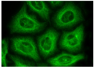
Immunofluorescence staining of methanol-fixed HeLa cells showing cytoplasmic and membrane localization.Application
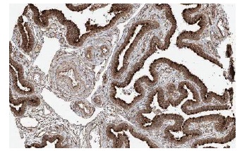
Immunoperoxidase staining of formalin fixed, paraffin-embedded human fallopian tube showing membrane staining of glandular cells at low magnification. Kindly provided by The Swedish Human Protein Atlas (HPA) program.Application
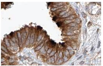
Immunoperoxidase staining of formalin fixed, paraffin-embedded human fallopian tube showing membrane staining of glandular cells at high magnification. Kindly provided by The Swedish Human Protein Atlas (HPA) program.Application
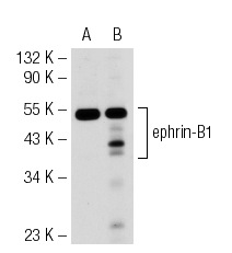
Western blot analysis of ephrin-B1 expression in non-transfected: (A) and human ephrin-B1 transfected: (B) 293T whole cell lysates.Application
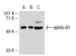
Western blot analysis of ephrin-B1 expression in non-transfected: (A) and mouse ephrin-B1 transfected: (B) 293T whole cell lysates and mouse embryo tissue extract (C).Application
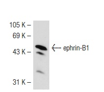
Western blot analysis of ephrin-B1 expression in human platelet whole cell lysate.| Product Name | ephrin-B1 Antibody (A-20) |
|---|---|
| Antibody Type | Primary Antibodies |
| Antigen Alias | epitope mapping within an internal region of ephrin-B1 of human origin |
| Modification Notes | Ephrins, which act as ligands for Eph receptors, are cell-surface proteins which fall into two categories, ephrin-A and ephrin-B, based on their structure and function. Ephrin-B proteins are transmembrane and have conserved cytoplasmic tyrosine residues that are phosphorylated upon interaction with an EphB receptor. Eph receptors and ephrins exhibit complementary expression in many tissues during embryogenesis indicating that bidirectional activation of Eph receptors and ephrin-B proteins may occur at expression domain interfaces. Ephrin-B1 transduces outside-in signals through C-terminal protein interactions that effect integrin-mediated cell attachment and migration. The distribution of ephrin-B1 in the developing retina suggests that it influences retinal axon mapping along the dorsal-ventral axis and may be involved in intratectal development. |
| Clonality | Polyclonal |
|---|---|
| Isotype | IgG |
| Host Species | Rabbit |
| Tested Applications | WBIPIFIHCELISA |
| recommended for detection of ephrin-B1 of mouse, rat and human origin by WB, IP, IF, IHC(P) and ELISA also reactive with additional species, including canine, bovine and avian: |
|
| Species Reactivity | HumanMouseRat |
| Concentration | 1mg/ml |
| Purification | Affinity purified |
| Alternative Names | epitope mapping within an internal region of ephrin-B1 of human origin |
|---|---|
| Tissue Specificity | epitope mapping within an internal region of ephrin-B1 of human origin |
| Entrez Gene | 1947 |
|---|
Application
Immunofluorescence staining of methanol-fixed HeLa cells showing cytoplasmic and membrane localization.
Application
Immunoperoxidase staining of formalin fixed, paraffin-embedded human fallopian tube showing membrane staining of glandular cells at low magnification. Kindly provided by The Swedish Human Protein Atlas (HPA) program.
Application
Immunoperoxidase staining of formalin fixed, paraffin-embedded human fallopian tube showing membrane staining of glandular cells at high magnification. Kindly provided by The Swedish Human Protein Atlas (HPA) program.
Application
Western blot analysis of ephrin-B1 expression in non-transfected: (A) and human ephrin-B1 transfected: (B) 293T whole cell lysates.
Application
Western blot analysis of ephrin-B1 expression in non-transfected: (A) and mouse ephrin-B1 transfected: (B) 293T whole cell lysates and mouse embryo tissue extract (C).
Application
Western blot analysis of ephrin-B1 expression in human platelet whole cell lysate.| Application Notes | recommended for detection of ephrin-B1 of mouse, rat and human origin by WB, IP, IF, IHC(P) and ELISA also reactive with additional species, including canine, bovine and avian: |
|---|
| Form | Liquid |
|---|---|
| Storage Instructions | For short-term storage, store at 4° C. For long-term storage, aliquot and store at -20ºC or below. Avoid multiple freeze-thaw cycles. |
| Storage Buffer | phosphate buffered saline , pH 7.4, 150mM NaCl, 0.02% sodium azide and 50% glycerol. |


