Application
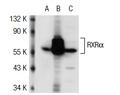
Western blot analysis of RXRα expression in non-transfected 293T: (A), human RXRα transfected 293T: (B) and K-562 (C) whole cell lysates.Application
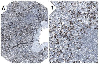
Immunoperoxidase staining of formalin fixed, paraffin-embedded human spleen tissue showing nuclear staining of cells in red pulp (low and high magnification). Kindly provided by The Swedish Human Protein Atlas (HPA) program.Application
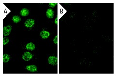
Immunofluorescence staining of methanol-fixed, control HeLa (A) and RXRα siRNA silenced HeLa (B) cells showing diminished nuclear staining in the siRNA silenced cells. Cells probed with RXRα (D-20): .Application
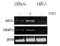
ChIP analysis of LXR dependent promoter recruitment of RXR in bone marrow-derived macrophages from LXR+/+ and LXR-/- mice in response to the LXR ligand T1317. Data kindly provided by M.G. Rosenfeld and reproduced with permission from Wagner et al., Mol. Cell. Biol. 2003, 23:5780-5789.Application
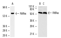
Antibodies tested include RARα (C-20): (A) and RXRα (D-20): (B,C).Application
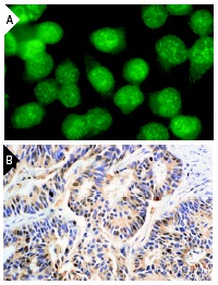
Immunofluorescence staining of methanol-fixed HeLa cells (A) and immunoperoxidase staining of formalin-fixed, paraffin-embedded human colon carcinoma tissue (B) showing nuclear staining.Application
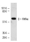
Western blot analysis of RXRα expression in HeLa nuclear extracts.| Product Name | RXRα Antibody (D-20) |
|---|---|
| Antibody Type | Primary Antibodies |
| Antigen Alias | epitope mapping at the N-terminus of RXRα of human origin |
| Modification Notes | Retinoids are metabolites of vitamin A (retinol) and are believed to represent important signaling molecules during vertebrate development and tissue differentiation. Two families of retinoid receptors have been identified. Retinoic acid receptors (RARs), include RARå RAR∫ and RAR©, each of which have a high affinity for all trans etinoic acids and belong to the same class of nuclear transcription factors as thyroid hormone receptors, vitamin D3 receptor and ecdysone receptor. The ligand binding domains of the RARs are highly conserved and RAR isoforms are expressed in distinct patterns throughout development and in the mature organism. Members of the retinoid X receptor (RXR) family, RXRå, RXR∫ and RXR©, are activated by 9-cis-RA, a stero- and photoisomer of all trans-RA, that is expressed in vivo in both liver and kidney and may represent a widely used hormone. As is true for the RAR subfamily, the RXR receptors are closely related to each other both in their DNA-binding and ligand-binding domains and are encoded by separate genes at distinct chromosomal loci. |
| Clonality | Polyclonal |
|---|---|
| Isotype | IgG |
| Host Species | Rabbit |
| Tested Applications | WBIPIFIHCELISAGSChIP |
| recommended for detection of RXRα of mouse, rat and human origin by WB, IP, IF, IHC(P) and ELISA: |
|
| Species Reactivity | HumanMouseRat |
| Concentration | 1mg/ml |
| Purification | Affinity purified |
| Alternative Names | epitope mapping at the N-terminus of RXR&alpha of human origin |
|---|---|
| Tissue Specificity | epitope mapping at the N-terminus of RXRα of human origin |
| Entrez Gene | 6256 |
|---|
Application
Western blot analysis of RXRα expression in non-transfected 293T: (A), human RXRα transfected 293T: (B) and K-562 (C) whole cell lysates.
Application
Immunoperoxidase staining of formalin fixed, paraffin-embedded human spleen tissue showing nuclear staining of cells in red pulp (low and high magnification). Kindly provided by The Swedish Human Protein Atlas (HPA) program.
Application
Immunofluorescence staining of methanol-fixed, control HeLa (A) and RXRα siRNA silenced HeLa (B) cells showing diminished nuclear staining in the siRNA silenced cells. Cells probed with RXRα (D-20): .
Application
ChIP analysis of LXR dependent promoter recruitment of RXR in bone marrow-derived macrophages from LXR+/+ and LXR-/- mice in response to the LXR ligand T1317. Data kindly provided by M.G. Rosenfeld and reproduced with permission from Wagner et al., Mol. Cell. Biol. 2003, 23:5780-5789.
Application
Antibodies tested include RARα (C-20): (A) and RXRα (D-20): (B,C).
Application
Immunofluorescence staining of methanol-fixed HeLa cells (A) and immunoperoxidase staining of formalin-fixed, paraffin-embedded human colon carcinoma tissue (B) showing nuclear staining.
Application
Western blot analysis of RXRα expression in HeLa nuclear extracts.| Application Notes | recommended for detection of RXRα of mouse, rat and human origin by WB, IP, IF, IHC(P) and ELISA: |
|---|
| Form | Liquid |
|---|---|
| Storage Instructions | For short-term storage, store at 4° C. For long-term storage, aliquot and store at -20ºC or below. Avoid multiple freeze-thaw cycles. |
| Storage Buffer | phosphate buffered saline , pH 7.4, 150mM NaCl, 0.02% sodium azide and 50% glycerol. |


