Application
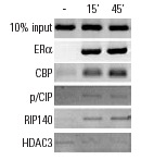
Antibodies tested include ERα (MC-20): , ERα (H-184): , CBP (A-22): , CBP (C-1): , p/CIP (F2): , p/CIP (M-397): , RIP140 (H-300): and HDAC3 (N-19): . Data kindly provided by M.G. Rosenfeld and reproduced with permission from Perissi et al., Cell 2004, 116: 511-526.Application
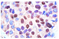
Immunoperoxidase staining of formalin-fixed, paraffin-embedded human breast carcinoma tissue showing nuclear localization.Application
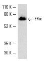
Western blot analysis of human recombinant ERα.IHC
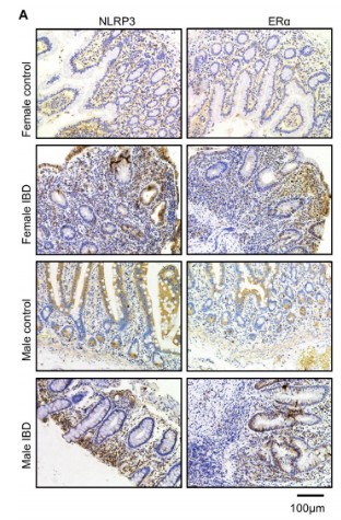
(A) Immunohistochemistry analysis of NLRP3 and ERα in different intestinal tissue sections. (Scale bars, 100µm).WB
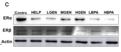
(C) Protein expression levels of ERα and ERβ in the liver tissue.WB
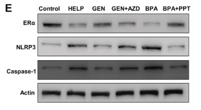
(E) Protein expression levels of ERα, NLRP3, and Caspase-1 of LMH cells.WB
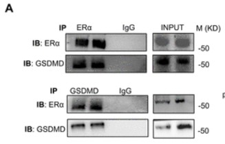
(A) Immunoblotting of immunoprecipitation with anti-ERα and anti-GSDMD in Hepa1-6 cells.WB
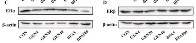
(C and D) Western blot analysis of ERα and ERβ.WB
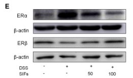
(E) Protein levels of ERα in the colon tissues. The relative protein expression of ERα was normalized to β-actin.WB
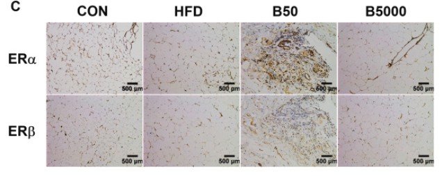
(C) ERα and ERβ in AFT were located by IHC. Bars: 500 µm.WB
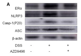
(A) Protein levels of NLRP3, ASC and Caspase-1 in colon tissues.WB
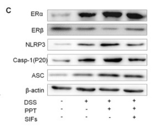
(C) Protein expression of NLRP3, ASC, Caspase-1 and ERα in colon tissues.IHC
(C) ERα and ERβ in AFT were located by IHC. Bars: 500 µm.WB
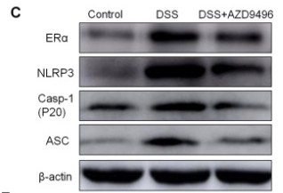
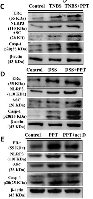
(C-E) The protein levels of ERα, NLRP3, ASC and Casp-1p20 in NCM-460 cells following different drug treatments.WB
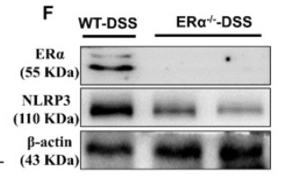
(F) The protein levels of ERα and NLRP3 in colon tissues.WB
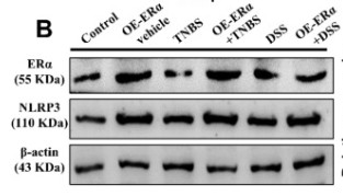
(B) The protein level of ERα and NLRP3 in ERα-overexpressing cells| Product Name | ERα Antibody (MC-20) |
|---|---|
| Antibody Type | Primary Antibodies |
| Conjugation | Agarose |
| Modification | Acetylated |
| Modification Notes | Estrogen receptors (ER) are members of the steroid/thyroid hormone receptor superfamily of ligand-activated transcription factors. Estrogen receptors, including ERå and ER∫, contain DNA binding and ligand binding domains and are critically involved in regulating the normal function of reproductive tissues. They are located in the nucleus , though some estrogen receptors associate with the cell surface membrane and can be rapidly activated by exposure of cells to estrogen. ERå and ER∫ have been shown to be differentially activated by various ligands. Receptor-ligand interactions trigger a cascade of events, including dissociation from heat shock proteins, receptor dimerization, phosphorylation and the association of the hormone activated receptor with specific regulatory elements in target genes. Evidence suggests that ERå and ER∫ may be regulated by distinct mechanisms even though they share many functional characteristics. |
| Clonality | Polyclonal |
|---|---|
| Isotype | IgG |
| Host Species | Rabbit |
| Tested Applications | ChIPELISAGSIFIHCIPWB |
| WB:1:100~1:500 IP:1:20~1:100 IF:1:20~1:100 IHC:1:10~1:50 |
|
| Species Reactivity | HumanMouseRat |
| Concentration | 1mg/ml |
| Purification | Affinity purified |
| Alternative Names | epitope mapping at the C-terminus of ERα of mouse origin |
|---|---|
| Tissue Specificity | epitope mapping at the C-terminus of ERα of mouse origin |
| Entrez Gene | 2099 |
|---|
Application
Antibodies tested include ERα (MC-20): , ERα (H-184): , CBP (A-22): , CBP (C-1): , p/CIP (F2): , p/CIP (M-397): , RIP140 (H-300): and HDAC3 (N-19): . Data kindly provided by M.G. Rosenfeld and reproduced with permission from Perissi et al., Cell 2004, 116: 511-526.
Application
Immunoperoxidase staining of formalin-fixed, paraffin-embedded human breast carcinoma tissue showing nuclear localization.
Application
Western blot analysis of human recombinant ERα.
IHC
(A) Immunohistochemistry analysis of NLRP3 and ERα in different intestinal tissue sections. (Scale bars, 100µm).
WB
(C) Protein expression levels of ERα and ERβ in the liver tissue.
WB
(E) Protein expression levels of ERα, NLRP3, and Caspase-1 of LMH cells.
WB
(A) Immunoblotting of immunoprecipitation with anti-ERα and anti-GSDMD in Hepa1-6 cells.
WB
(C and D) Western blot analysis of ERα and ERβ.
WB
(E) Protein levels of ERα in the colon tissues. The relative protein expression of ERα was normalized to β-actin.
WB
(C) ERα and ERβ in AFT were located by IHC. Bars: 500 µm.
WB
(A) Protein levels of NLRP3, ASC and Caspase-1 in colon tissues.
WB
(C) Protein expression of NLRP3, ASC, Caspase-1 and ERα in colon tissues.
IHC
(C) ERα and ERβ in AFT were located by IHC. Bars: 500 µm.
WB
(C-E) The protein levels of ERα, NLRP3, ASC and Casp-1p20 in NCM-460 cells following different drug treatments.
WB
(F) The protein levels of ERα and NLRP3 in colon tissues.
WB
(B) The protein level of ERα and NLRP3 in ERα-overexpressing cells| Application Notes | WB:1:100~1:500 IP:1:20~1:100 IF:1:20~1:100 IHC:1:10~1:50 |
|---|
| Form | Liquid |
|---|---|
| Storage Instructions | For short-term storage, store at 4° C. For long-term storage, aliquot and store at -20ºC or below. Avoid multiple freeze-thaw cycles. |
| Storage Buffer | phosphate buffered saline , pH 7.4, 150mM NaCl, 0.02% sodium azide and 50% glycerol. |


