
IF
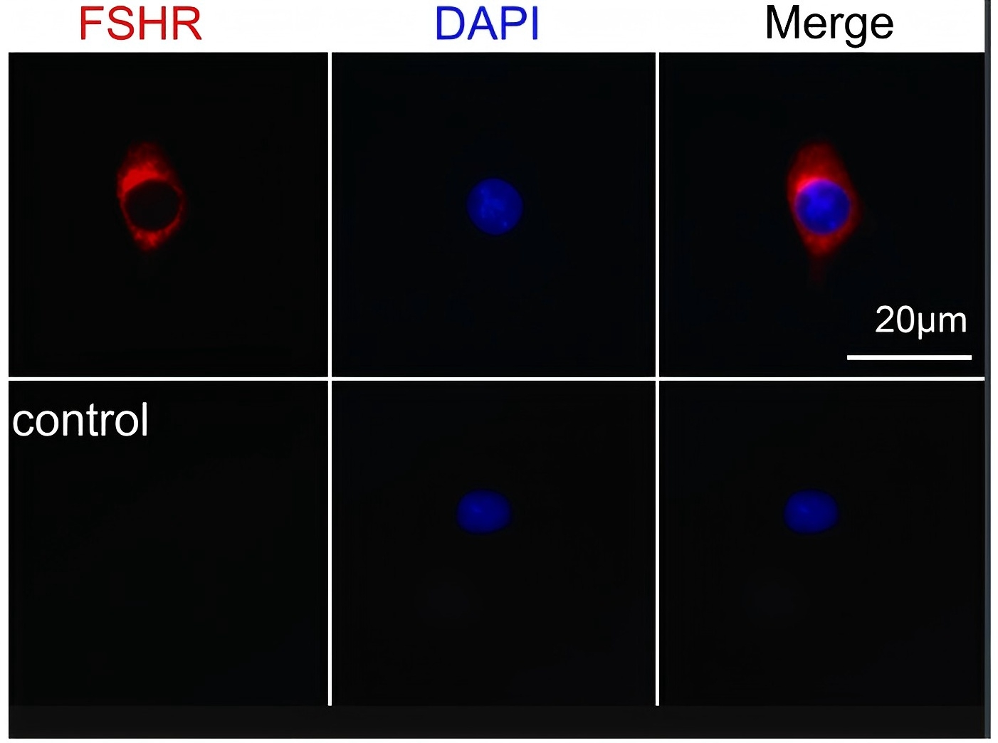
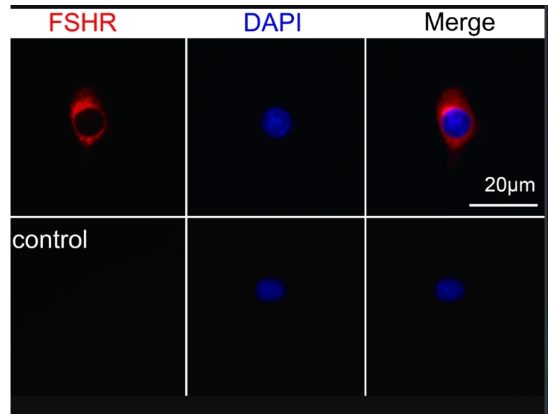
we used FSH to pretreat N2a cells, a mouse neuroblastoma cell line expressing FSHR (Fig. S3), and then induced cellular oxidative stress by the addition of hydrogen peroxide to observe the protective effect of FSH on cellular oxidative stress.IF
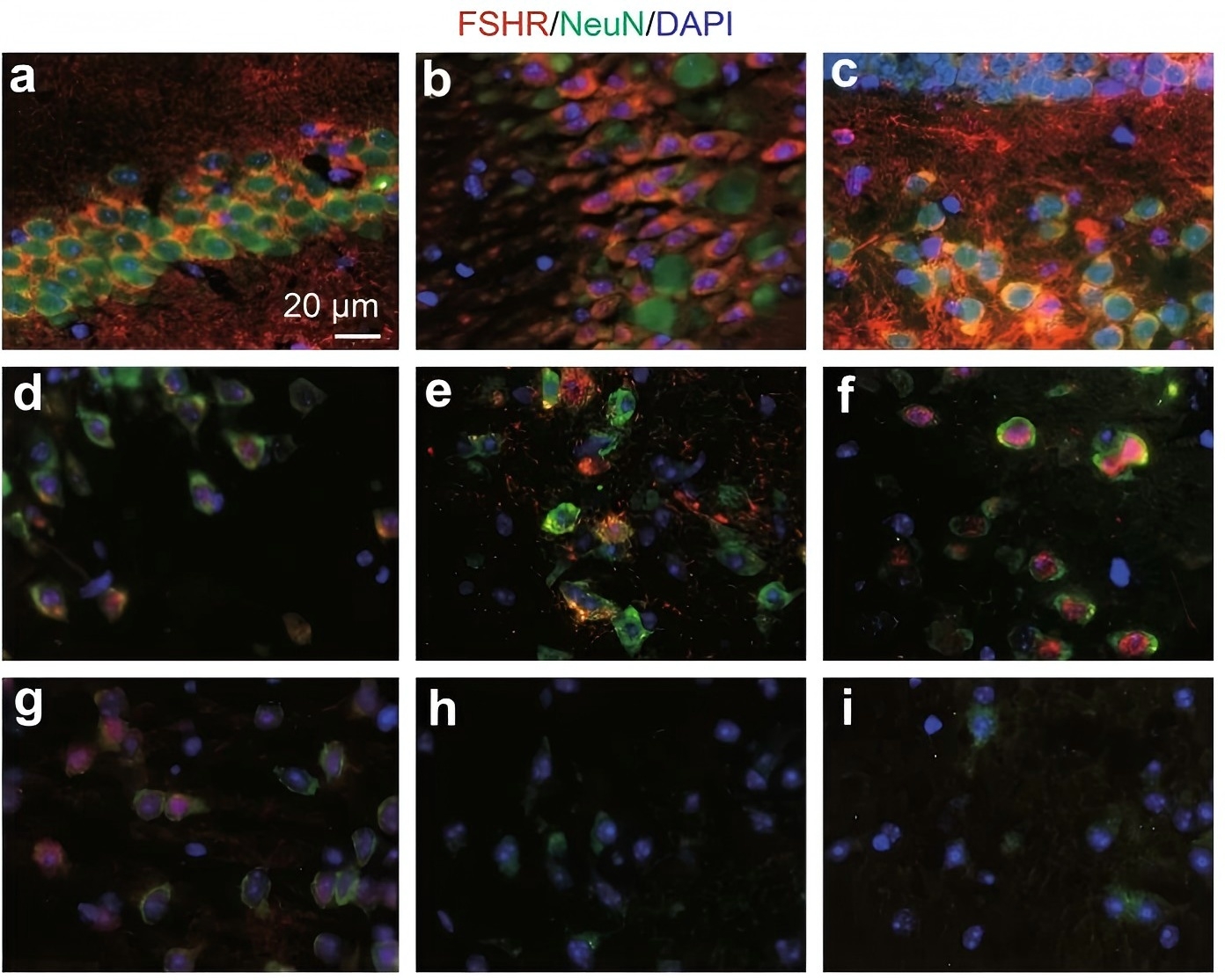
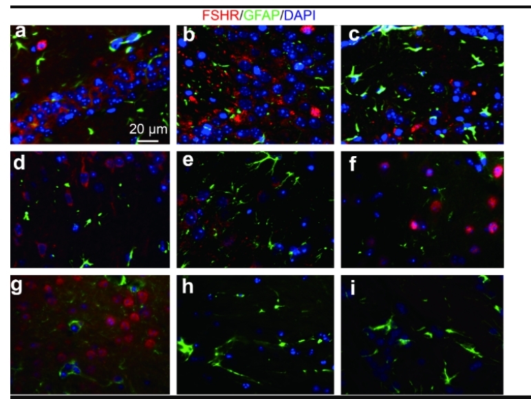
The expression of FSHR in depression-related brain regions. a The CA1 region of the mouse hippocampus. b The CA3 region of mouse hippocampus. c The DG of the mouse hippocampus. d The cortex. e The nucleus accumbens. f The amygdala. g The prefrontal cortex. h The bed nucleus of the stria terminalis. i The lateral habenula. FSHR (red), NeuN (green, a marker of neurons), and DAPI (blue) immunofluorescence was detected under a fluorescence microscopeIHC
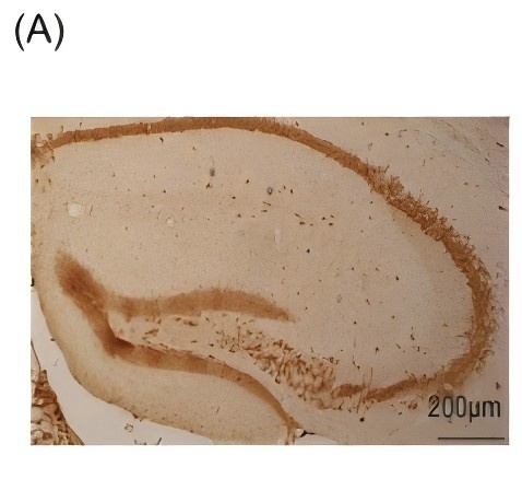
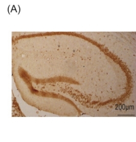
(A) Immunohistochemical stain of FSHR in mouse hippocampus.IHC
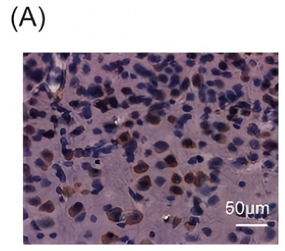
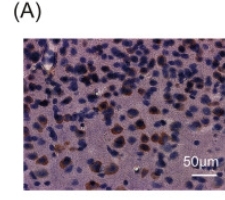
(A) Immunohistochemical staining of FSHR in the prefrontal cortex.IF
The expression of FSHR in depression-related brain regions. a The CA1 region of the mouse hippocampus. b The CA3 region of mouse hippocampus. c The DG of the mouse hippocampus. d The cortex. e The nucleus accumbens. f The amygdala. g The prefrontal cortex. h The bed nucleus of the stria terminalis. i The lateral habenula. FSHR (red), NeuN (green, a marker of neurons), and DAPI (blue) immunofluorescence was detected under a fluorescence microscopeIF
The immunofluorescence results showed that FSHR was present in neurons, but not astrocytes (Fig. S1), in multiple depression-related regions, including the hippocampus, cortex, nucleus accumbens, amygdala, and prefrontal cortex (Fig. 1a–g), not in the bed nucleus of the stria terminalis or lateral habenula (Fig. 1h, i). PreviousIF
Next, we used FSH to pretreat N2a cells, a mouse neuroblastoma cell line expressing FSHR (Fig. S3), and then induced cellular oxidative stress by the addition of hydrogen peroxide to observe the protective effect of FSH on cellular oxidative stress. PreviousIHC
(A) Immunohistochemical stain of FSHR in mouse hippocampus. PreviousIHC
(A) Immunohistochemical staining of FSHR in the prefrontal cortex. Previous| Product Name | FSHR Antibody (H-190) |
|---|---|
| Antibody Type | Primary Antibodies |
| Conjugation | Agarose |
| Modification | Acetylated |
| Modification Notes | Follicle-stimulating hormone receptor (FSHR) is a 695 amino acid G-protein coupled receptor. FSH binds to the receptor in a hand-clasp fashion via its å and ∫ subunits. While the å subunit of FSH is involved in the binding of FSH to the receptor, the ∫ subunit stabilizes this interaction. Linkage studies suggest that a missense mutation in the FSHR gene can cause reduced FSH binding affinity and lead to a condition known as hypergonadotropic ovarian dysgenesis (ODG). In males however, this mutation does not appear to have a detrimental affect on fertility. It is believed that a mutation in the FSHR gene is also associated with ovarian hyperstimulation syndrome; a condition characterized by the presence of multiple serous and hemorrhagic follicular cysts lined by luteinized cells. |
| Clonality | Polyclonal |
|---|---|
| Isotype | IgG |
| Host Species | Rabbit |
| Tested Applications | ELISAIFIHCIPWB |
| recommended for detection of FSHR of mouse, rat and human origin by WB, IP, IF and ELISA also reactive with additional species, including canine: |
|
| Species Reactivity | HumanMouseRat |
| Concentration | 1mg/ml |
| Purification | Affinity purified |
| Alternative Names | epitope corresponding to amino acids 1-190 mapping at the N-terminus of follicle-stimulating hormone receptor (FSHR) of human origin |
|---|---|
| Tissue Specificity | epitope corresponding to amino acids 1-190 mapping at the N-terminus of follicle-stimulating hormone receptor (FSHR) of human origin |
| Entrez Gene | 2492 |
|---|
IF
we used FSH to pretreat N2a cells, a mouse neuroblastoma cell line expressing FSHR (Fig. S3), and then induced cellular oxidative stress by the addition of hydrogen peroxide to observe the protective effect of FSH on cellular oxidative stress.
IF
The expression of FSHR in depression-related brain regions. a The CA1 region of the mouse hippocampus. b The CA3 region of mouse hippocampus. c The DG of the mouse hippocampus. d The cortex. e The nucleus accumbens. f The amygdala. g The prefrontal cortex. h The bed nucleus of the stria terminalis. i The lateral habenula. FSHR (red), NeuN (green, a marker of neurons), and DAPI (blue) immunofluorescence was detected under a fluorescence microscope
IHC
(A) Immunohistochemical stain of FSHR in mouse hippocampus.
IHC
(A) Immunohistochemical staining of FSHR in the prefrontal cortex.
IF
The expression of FSHR in depression-related brain regions. a The CA1 region of the mouse hippocampus. b The CA3 region of mouse hippocampus. c The DG of the mouse hippocampus. d The cortex. e The nucleus accumbens. f The amygdala. g The prefrontal cortex. h The bed nucleus of the stria terminalis. i The lateral habenula. FSHR (red), NeuN (green, a marker of neurons), and DAPI (blue) immunofluorescence was detected under a fluorescence microscope
IF
The immunofluorescence results showed that FSHR was present in neurons, but not astrocytes (Fig. S1), in multiple depression-related regions, including the hippocampus, cortex, nucleus accumbens, amygdala, and prefrontal cortex (Fig. 1a–g), not in the bed nucleus of the stria terminalis or lateral habenula (Fig. 1h, i). Previous
IF
Next, we used FSH to pretreat N2a cells, a mouse neuroblastoma cell line expressing FSHR (Fig. S3), and then induced cellular oxidative stress by the addition of hydrogen peroxide to observe the protective effect of FSH on cellular oxidative stress. Previous
IHC
(A) Immunohistochemical stain of FSHR in mouse hippocampus. Previous
IHC
(A) Immunohistochemical staining of FSHR in the prefrontal cortex. Previous| Application Notes | recommended for detection of FSHR of mouse, rat and human origin by WB, IP, IF and ELISA also reactive with additional species, including canine: |
|---|
| Form | Liquid |
|---|---|
| Storage Instructions | For short-term storage, store at 4° C. For long-term storage, aliquot and store at -20ºC or below. Avoid multiple freeze-thaw cycles. |
| Storage Buffer | phosphate buffered saline , pH 7.4, 150mM NaCl, 0.02% sodium azide and 50% glycerol. |


