Application
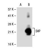
Western blot analysis of SHP expression in non-transfected: (A) and human SHP transfected: (B) 293T whole cell lysates.IF
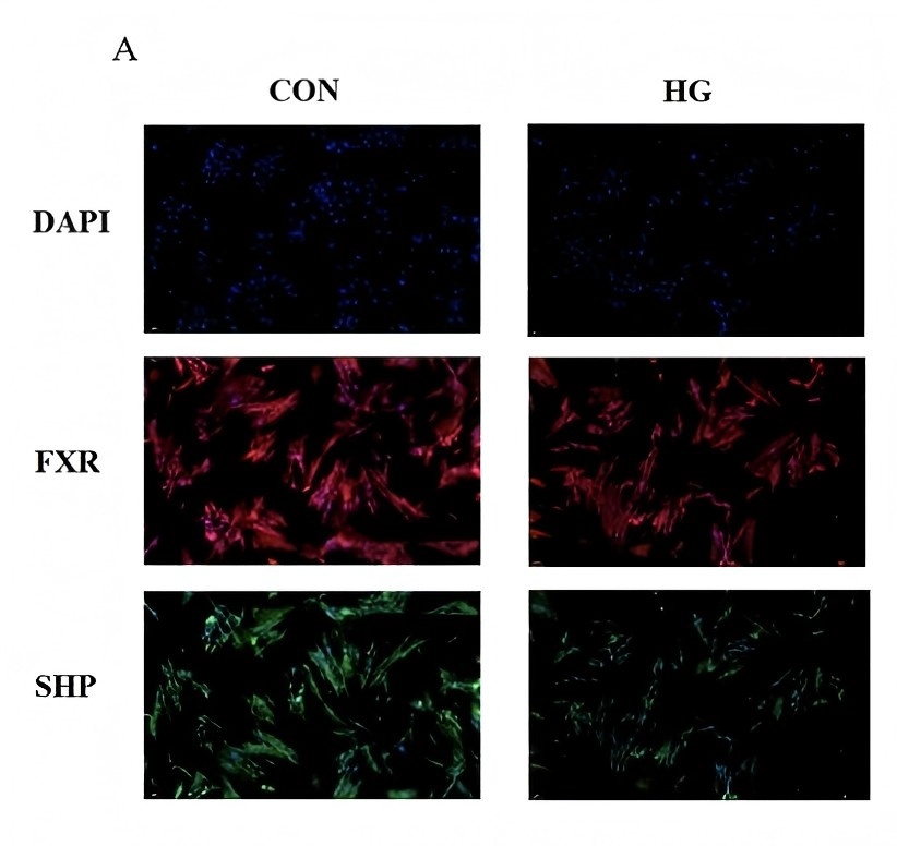
(A) Immunofluorescence results of FXR and SHP at different glucose levels. Red fluorescence represents the expression of FXR, green fluorescence represents the expression of SHP, and blue represents the nucleus stained with DAPI.| Product Name | SHP Antibody (H-160) |
|---|---|
| Antibody Type | Primary Antibodies |
| Modification Notes | SHP (short heterodimer partner and small heterodimer partner) is an orphan nuclear receptor containing the dimerization and ligand-binding domains found in other nuclear receptors, but lacking the conserved DNA binding domain. SHP is specifically expressed in liver and other tissues, including fetal liver and adrenal gland, as well as adult spleen and small intestine. In addition, SHP is highy expressed in the murine macrophage cell line RAW 264.7 but suppressed by oxLDL and 13-HODE, which is a ligand for PPARg. SHP interacts with nuclear receptors, including thyroid receptor, retinoic acid receptors (RAR and RXR) and estrogen receptors (ERa and ERb). SHP functions as a negative regulator of these receptors by at least three mechanisms: inhibition of DNA binding via dimerization, direct antagonism of coactivator function through competition and possibly transrepression via recruitment of putative corepressors. In oxLDL-treated, resting macrophage cells, SHP acts as a transcription coactivator of NFkB, suggesting that SHP is a modulatory component in the regulation of the transcriptional activities of NFkB. Lastly, negative feedback regulation of a hepatic bile acid transporter, NTCP, is controlled by bile acid-activated FXR via induction of SHP to protect the hepatocyte from bile acid-mediated damage in cholestatic conditions. |
| Clonality | Polyclonal |
|---|---|
| Isotype | IgG |
| Host Species | Rabbit |
| Tested Applications | ELISAIFIPWB |
| recommended for detection of SHP of mouse, rat and human origin by WB, IP, IF and ELISA also reactive with additional species, including canine | |
| Species Reactivity | HumanMouseRat |
| Concentration | 1mg/ml |
| Purification | Affinity purified |
| Alternative Names | epitope corresponding to amino acids 1-160 mapping at the N-terminus of SHP of human origin |
|---|---|
| Tissue Specificity | epitope corresponding to amino acids 1-160 mapping at the N-terminus of SHP of human origin |
| Entrez Gene | 8431 |
|---|
Application
Western blot analysis of SHP expression in non-transfected: (A) and human SHP transfected: (B) 293T whole cell lysates.
IF
(A) Immunofluorescence results of FXR and SHP at different glucose levels. Red fluorescence represents the expression of FXR, green fluorescence represents the expression of SHP, and blue represents the nucleus stained with DAPI.| Application Notes | recommended for detection of SHP of mouse, rat and human origin by WB, IP, IF and ELISA also reactive with additional species, including canine |
|---|
| Form | Liquid |
|---|---|
| Storage Instructions | For short-term storage, store at 4° C. For long-term storage, aliquot and store at -20ºC or below. Avoid multiple freeze-thaw cycles. |
| Storage Buffer | phosphate buffered saline , pH 7.4, 150mM NaCl, 0.02% sodium azide and 50% glycerol. |


