Application
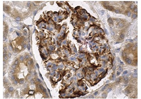
Immunoperoxidase staining of formalin fixed, paraffin-embedded human kidney tissue showing membrane staining of glomerular cells and cytoplasmic staining of cells in tubules.Application
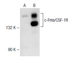
Western blot analysis of c-Fms/CSF-1R expression in non-transfected: (A) and mouse c-Fms/CSF-1R transfected: (B) 293T whole cell lysates.Application
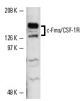
Western blot analysis of c-Fms/CSF-1R expression in RAW 264.7 whole cell lysate. The two major bands represent differentially glycosylated receptors.| Product Name | c-Fms/CSF-1R Antibody (H-300) |
|---|---|
| Antibody Type | Primary Antibodies |
| Modification Notes | c-Fms/CSF-1R, also designated macrophage colony-stimulating factor receptor (M-CSFR), FIM2 or CD115, is a transmembrane tyrosine kinase receptor belonging to the CSF1/PDGF receptor family. It is encoded by the c-Fms proto-oncogene and is expressed in mononuclear phagocytes, oocytes, decidual cells, trophoblastic cells and some myoblasts. It is important for growth and differentiation of myeloid cells and its function can be regulated by SLAP-2. c-Fms/CSF-1R is responsible for mediating all of the functions of M-CSF. M-CSF is a glycoprotein required for the proliferation and differentiation of mononuclear phagocytes, including osteoclasts. M-CSF has also been identified as an important mediator of the inflammatory response and can regulate the release of proinflammatory cytokines from macrophages. |
| Clonality | Polyclonal |
|---|---|
| Isotype | IgG |
| Host Species | Rabbit |
| Tested Applications | WBIPIFIHCELISA |
| recommended for detection of c-Fms/CSF-1R of mouse, rat and human origin by WB, IP, IF, IHC(P) and ELISA | |
| Species Reactivity | HumanMouseRat |
| Concentration | 1mg/ml |
| Purification | Affinity purified |
| Alternative Names | epitope corresponding to amino acids 11-310 mapping near the N-terminus of c-Fms/CSF-1R of human origin |
|---|---|
| Tissue Specificity | epitope corresponding to amino acids 11-310 mapping near the N-terminus of c-Fms/CSF-1R of human origin |
| Entrez Gene | 1436 |
|---|
Application
Immunoperoxidase staining of formalin fixed, paraffin-embedded human kidney tissue showing membrane staining of glomerular cells and cytoplasmic staining of cells in tubules.
Application
Western blot analysis of c-Fms/CSF-1R expression in non-transfected: (A) and mouse c-Fms/CSF-1R transfected: (B) 293T whole cell lysates.
Application
Western blot analysis of c-Fms/CSF-1R expression in RAW 264.7 whole cell lysate. The two major bands represent differentially glycosylated receptors.| Application Notes | recommended for detection of c-Fms/CSF-1R of mouse, rat and human origin by WB, IP, IF, IHC(P) and ELISA |
|---|
| Form | Liquid |
|---|---|
| Storage Instructions | For short-term storage, store at 4° C. For long-term storage, aliquot and store at -20ºC or below. Avoid multiple freeze-thaw cycles. |
| Storage Buffer | phosphate buffered saline , pH 7.4, 150mM NaCl, 0.02% sodium azide and 50% glycerol. |


