Application
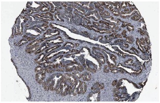
Immunoperoxidase staining of formalin fixed, paraffin-embedded human seminal vesicle tissue showing cytoplasmic staining of glandular cells at low magnification. Kindly provided by The Swedish Human Protein Atlas (HPA) program.Application
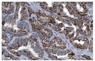
Immunoperoxidase staining of formalin fixed, paraffin-embedded human seminal vesicle tissue showing cytoplasmic staining of glandular cells at high magnification. Kindly provided by The Swedish Human Protein Atlas (HPA) program.Application
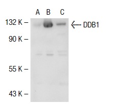
Western blot analysis of DDB1 expression in non-transfected 293T: (A), mouse DDB1 transfected 293T: (B) and UV HeLa (C) whole cell lysates.Application
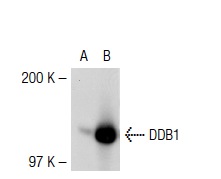
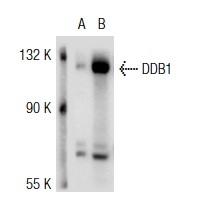
Western blot analysis of DDB1 expression in non-transfected: (A) and human DDB1 transfected: (B) 293T whole cell lysates.Application
Western blot analysis of DDB1 expression in non-transfected: (A) and human DDB1 transfected: (B) 293T whole cell lysates.| Product Name | DDB1 Antibody (H-300) |
|---|---|
| Antibody Type | Primary Antibodies |
| Modification Notes | Damaged DNA binding protein (DDB) is a heterodimer composed of two subunits, p127 and p48, which are designated DDB1 and DDB2, respectively. The DDB heterodimer is involved in repairing DNA damaged by ultraviolet light. Specifically, DDB, also designated UV-damaged DNA binding protein (UV-DDB), xeroderma pigmentosum group E binding factor (XPE-BF) and hepatitis B virus X-associated protein 1 (XAP-1), binds to damaged cyclobutane pyrimidine dimers (CPDs). Mutations in the DDB2 gene are implicated as causes of xeroderma pigmentosum group E, an autosomal recessive disease in which patients are defective in nucleotide excision DNA repair. XPE is characterized by hypersensitivity of the skin to sunlight with a high frequency of skin cancer as well as neurologic abnormalities. The hepatitis B virus (HBV) X protein interacts with DDB1, which may mediate HBx transactivation. |
| Clonality | Polyclonal |
|---|---|
| Isotype | IgG |
| Host Species | Rabbit |
| Tested Applications | WBIPIFIHCELISA |
| recommended for detection of DDB1 of mouse, rat and human origin by WB, IP, IF, IHC(P) and ELISA | |
| Species Reactivity | HumanMouseRat |
| Concentration | 1mg/ml |
| Purification | Affinity purified |
| Alternative Names | epitope corresponding to amino acids 1-300 of DDB1 of human origin |
|---|---|
| Tissue Specificity | epitope corresponding to amino acids 1-300 of DDB1 of human origin |
| Entrez Gene | 1642 |
|---|
Application
Immunoperoxidase staining of formalin fixed, paraffin-embedded human seminal vesicle tissue showing cytoplasmic staining of glandular cells at low magnification. Kindly provided by The Swedish Human Protein Atlas (HPA) program.
Application
Immunoperoxidase staining of formalin fixed, paraffin-embedded human seminal vesicle tissue showing cytoplasmic staining of glandular cells at high magnification. Kindly provided by The Swedish Human Protein Atlas (HPA) program.
Application
Western blot analysis of DDB1 expression in non-transfected 293T: (A), mouse DDB1 transfected 293T: (B) and UV HeLa (C) whole cell lysates.
Application
Western blot analysis of DDB1 expression in non-transfected: (A) and human DDB1 transfected: (B) 293T whole cell lysates.
Application
Western blot analysis of DDB1 expression in non-transfected: (A) and human DDB1 transfected: (B) 293T whole cell lysates.| Application Notes | recommended for detection of DDB1 of mouse, rat and human origin by WB, IP, IF, IHC(P) and ELISA |
|---|
| Form | Liquid |
|---|---|
| Storage Instructions | For short-term storage, store at 4° C. For long-term storage, aliquot and store at -20ºC or below. Avoid multiple freeze-thaw cycles. |
| Storage Buffer | phosphate buffered saline , pH 7.4, 150mM NaCl, 0.02% sodium azide and 50% glycerol. |


