Application
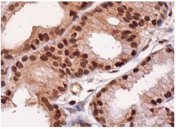
Immunoperoxidase staining of formalin fixed, paraffin-embedded human salivary gland tissue showing cytoplasmic and nuclear staining of glandular cells.Application
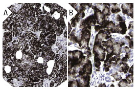
Immunoperoxidase staining of formalin fixed, paraffin-embedded human pancreas tissue showing cytoplasmic staining in exocrine pancreas at low (A) and high (B) magnification. Kindly provided by The Swedish Human Protein Atlas (HPA) program.Application
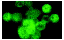
Immunofluorescence staining of methanol-fixed HeLa cells showing cytoplasmic localization.Application
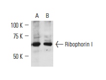
Western blot analysis of Ribophorin I expression in KNRK (A) and NIH/3T3 (B) whole cell lysates.| Product Name | Ribophorin I Antibody (H-50) |
|---|---|
| Antibody Type | Primary Antibodies |
| Modification Notes | Membrane proteins of the endoplasmic reticulum (ER) may be localized by mechanisms that involve retention, retrieval or a combination of both. ER localization information has been found in cytoplasmic, transmembrane or luminal domains. Specific retrieval mechanisms have been identified for luminal ER proteins, which contain a KDEL domain, and for type I transmembrane proteins carrying a dilysine motif. Mammalian oligosaccharyltransferase (OST) is a protein complex that is composed of four rough ER-specific, type I transmembrane proteins: Ribophorins I and II (RI and RII), OST48 and DAD1 (also designated defender against apoptotic death). The ribophorins are integral membrane glycoproteins that localize exclusively to the rough ER. There is affinity between the cytoplasmically located N-terminal region of DAD1 and the short cytoplasmic tail of OST48 to place DAD1 firmly into the OST complex. The OST complex affects the cotranslational N-glycosylation of newly synthesized polypeptides. |
| Clonality | Polyclonal |
|---|---|
| Isotype | IgG |
| Host Species | Rabbit |
| Tested Applications | WB,IP,IF,IHC,ELISA |
| recommended for detection of Ribophorin I of mouse, rat and human origin by WB, IP, IF, IHC(P) and ELISA also reactive with additional species, including equine, canine, bovine, porcine and avian | |
| Species Reactivity | Human,Mouse,Rat |
| Concentration | 1mg/ml |
| Purification | Affinity purified |
| Alternative Names | epitope corresponding to amino acids 558-607 of Ribophorin I of human origin |
|---|---|
| Tissue Specificity | epitope corresponding to amino acids 558-607 of Ribophorin I of human origin |
| Entrez Gene | 6184 |
|---|
Application
Immunoperoxidase staining of formalin fixed, paraffin-embedded human salivary gland tissue showing cytoplasmic and nuclear staining of glandular cells.
Application
Immunoperoxidase staining of formalin fixed, paraffin-embedded human pancreas tissue showing cytoplasmic staining in exocrine pancreas at low (A) and high (B) magnification. Kindly provided by The Swedish Human Protein Atlas (HPA) program.
Application
Immunofluorescence staining of methanol-fixed HeLa cells showing cytoplasmic localization.
Application
Western blot analysis of Ribophorin I expression in KNRK (A) and NIH/3T3 (B) whole cell lysates.| Application Notes | recommended for detection of Ribophorin I of mouse, rat and human origin by WB, IP, IF, IHC(P) and ELISA also reactive with additional species, including equine, canine, bovine, porcine and avian |
|---|
| Form | Liquid |
|---|---|
| Storage Instructions | For short-term storage, store at 4° C. For long-term storage, aliquot and store at -20ºC or below. Avoid multiple freeze-thaw cycles. |
| Storage Buffer | phosphate buffered saline , pH 7.4, 150mM NaCl, 0.02% sodium azide and 50% glycerol. |


