Application
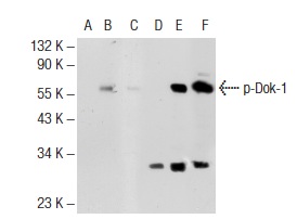
Antibodies tested include p-Dok-1 (Tyr 362): (A, B, C) and Dok-1 (M-276): (D, E, F).Application
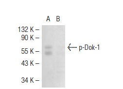
Western blot analysis of Dok-1 phosphorylation in untreated (A) and lambda protein phosphatase treated (B) Jurkat whole cell lysates.Application
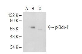
Western blot analysis of Dok-1 phosphorylation in non-transfected: (A), untreated mouse Dok-1 transfected: (B) and lambda protein phosphatase treated mouse Dok-1 transfected: (C) 293T whole cell lysates.Application
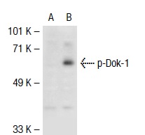
Western blot analysis of Dok-1 phosphorylation in non-transfected: (A) and mouse Dok-1 transfected: (B) 293T whole cell lysates.Application
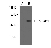
Blots were probed with p-Dok-1 (Tyr 362): : preincubated with its cognate phosphorylated peptide (A) and p-Dok-1 (Tyr 362): (B).Application
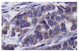
Immunoperoxidase staining of formalin-fixed, paraffin-embedded human breast carcinoma tissue showing nuclear and cytoplasmic staining.| Product Name | p-Dok-1 Antibody (Tyr 362) |
|---|---|
| Antibody Type | Primary Antibodies |
| Antigen Alias | epitope corresponding to phosphorylated Tyr 362 of Dok-1 of human origin |
| Modification | Phosphoralated |
| Modification Notes | Dok-1 associates with the Ras GTPase activating protein (Ras GAP) upon tyrosine phosphorylation. Evidence suggests that p62 Dok-1 is a substrate of the constitutive tyrosine kinase activity of p210 Bcr-Abl, a fusion protein caused by the t(9;22) translocation and associated with chronic myelogenous leukemia. Dok-1, as well as the tyrosine kinase substrates IRS-1 and Cas, is a member of a class of “docking” proteins which contain multiple tyrosine residues and putative SH2 binding sites. Dok-1 is suspected to be the substrate phosphorylated in response to stimulation by a number of growth factors, including PDGF, VEGF, insulin and IGF. Dok-2 (also designated p56 Dok) has also been identified as a potential mediator of the effects of p210 Bcr-Abl. |
| Clonality | Polyclonal |
|---|---|
| Isotype | IgG |
| Host Species | Rabbit |
| Tested Applications | WBIPIFIHC |
| recommended for detection of Tyr 362 phosphorylated Dok-1 of mouse, rat and human origin by WB, IP, IF and IHC(P): |
|
| Species Reactivity | HumanMouseRat |
| Concentration | 1mg/ml |
| Purification | Affinity purified |
| Alternative Names | epitope corresponding to phosphorylated Tyr 362 of Dok-1 of human origin |
|---|---|
| Tissue Specificity | epitope corresponding to phosphorylated Tyr 362 of Dok-1 of human origin |
| Entrez Gene | 1796 |
|---|
Application
Antibodies tested include p-Dok-1 (Tyr 362): (A, B, C) and Dok-1 (M-276): (D, E, F).
Application
Western blot analysis of Dok-1 phosphorylation in untreated (A) and lambda protein phosphatase treated (B) Jurkat whole cell lysates.
Application
Western blot analysis of Dok-1 phosphorylation in non-transfected: (A), untreated mouse Dok-1 transfected: (B) and lambda protein phosphatase treated mouse Dok-1 transfected: (C) 293T whole cell lysates.
Application
Western blot analysis of Dok-1 phosphorylation in non-transfected: (A) and mouse Dok-1 transfected: (B) 293T whole cell lysates.
Application
Blots were probed with p-Dok-1 (Tyr 362): : preincubated with its cognate phosphorylated peptide (A) and p-Dok-1 (Tyr 362): (B).
Application
Immunoperoxidase staining of formalin-fixed, paraffin-embedded human breast carcinoma tissue showing nuclear and cytoplasmic staining.| Application Notes | recommended for detection of Tyr 362 phosphorylated Dok-1 of mouse, rat and human origin by WB, IP, IF and IHC(P): |
|---|
| Form | Liquid |
|---|---|
| Storage Instructions | For short-term storage, store at 4° C. For long-term storage, aliquot and store at -20ºC or below. Avoid multiple freeze-thaw cycles. |
| Storage Buffer | phosphate buffered saline , pH 7.4, 150mM NaCl, 0.02% sodium azide and 50% glycerol. |


