Application
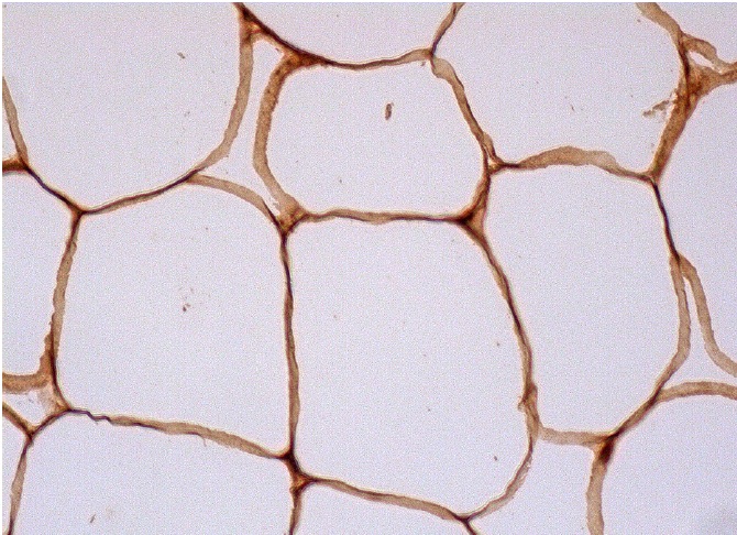
Immunoperoxidase staining of formalin fixed, paraffin-embedded human appendix tissue showing membrane staining of adipocytes.Application
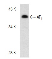
Western blot analysis of AT1 expression in A549 whole cell lysate.Application
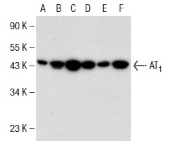
Western blot analysis of AT1 expression in c4 (A), NIH/3T3 (B), C3H/10T1/2 (C), A549 (D), Mv 1 Lu (E) and A-10 (F) whole cell lysates.Application
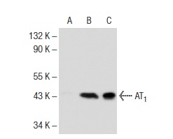
Western blot analysis of AT1 expression in non-transfected: (A) and human AT1 transfected: (B) 293T whole cell lysates and rat brain tissue extract (C).Application
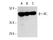
Western blot analysis of AT1 expression in c4 (A), NIH/3T3 (B) and C3H/10T1/2 (C) whole cell lysates.Application
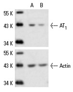
Western blot analysis of AT1 expression in non-transfected control (A) and AT1 siRNA transfected (B) HeLa cells. Blot probed with AT1 (N-10): . Actin (I-19): used as specificity and loading control.| Product Name | AT1 Antibody (N-10) AC |
|---|---|
| Antibody Type | Primary Antibodies |
| Modification Notes | Angiotensin II (Ang II) is an important physiological effector of blood pressure and volume regulation through vasoconstriction, aldosterone release, sodium uptake and thirst stimulation. Although Ang II interacts with two types of cell surface receptors, AT1 and AT2, most of the major cardiovascular effects seem to be mediated through AT1. Molecular cloning of the AT1 protein has shown it to be a member of the G protein-associated seven transmembrane protein receptor family. Ang II treatment of cells results in activation of several signal transduction pathways as evidenced by tyrosine phosphorylation of several proteins and induction of others. PLC© is phosphorylated after 30 seconds of treatment with Angiotensin II, indicating this as an early signal transduction event. Ang II treatment also stimulates phosphorylation of Shc, FAK and MAP kinases, and induces MKP-1, indicating stimulation of growth factor pathways. Ang II stimulation through AT1 has been shown to activate the JAK/Stat pathway involving a direct interaction between JAK2 and AT1 as demonstrated by coimmunoprecipitation. The AT1 receptor has no cytoplasmic kinase domain, but is able to function as a substrate for Src kinases and has several putative phosphorylation sites. |
| Clonality | Polyclonal |
|---|---|
| Isotype | IgG |
| Host Species | Rabbit |
| Tested Applications | WB,IP,IF,ELISA |
| recommended for detection of AT1 of mouse, rat and human origin by WB, IP, IF and ELISA also reactive with additional species, including equine, canine, bovine and porcine | |
| Species Reactivity | Human,Mouse,Rat |
| Concentration | 1mg/ml |
| Purification | Affinity purified |
| Alternative Names | epitope mapping within an N-terminal extracellular domain of AT1 of human origin |
|---|---|
| Tissue Specificity | epitope mapping within an N-terminal extracellular domain of AT1 of human origin |
| Entrez Gene | 185 |
|---|
Application
Immunoperoxidase staining of formalin fixed, paraffin-embedded human appendix tissue showing membrane staining of adipocytes.
Application
Western blot analysis of AT1 expression in A549 whole cell lysate.
Application
Western blot analysis of AT1 expression in c4 (A), NIH/3T3 (B), C3H/10T1/2 (C), A549 (D), Mv 1 Lu (E) and A-10 (F) whole cell lysates.
Application
Western blot analysis of AT1 expression in non-transfected: (A) and human AT1 transfected: (B) 293T whole cell lysates and rat brain tissue extract (C).
Application
Western blot analysis of AT1 expression in c4 (A), NIH/3T3 (B) and C3H/10T1/2 (C) whole cell lysates.
Application
Western blot analysis of AT1 expression in non-transfected control (A) and AT1 siRNA transfected (B) HeLa cells. Blot probed with AT1 (N-10): . Actin (I-19): used as specificity and loading control.| Application Notes | recommended for detection of AT1 of mouse, rat and human origin by WB, IP, IF and ELISA also reactive with additional species, including equine, canine, bovine and porcine |
|---|
| Form | Liquid |
|---|---|
| Storage Instructions | For short-term storage, store at 4° C. For long-term storage, aliquot and store at -20ºC or below. Avoid multiple freeze-thaw cycles. |
| Storage Buffer | phosphate buffered saline , pH 7.4, 150mM NaCl, 0.02% sodium azide and 50% glycerol. |


