Application
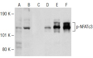
Antibodies tested include p-NFATc3 (Ser 240): (A,B,C) and NFATc3 (F-1): (D,E,F).| Product Name | p-NFATc3 Antibody (Ser 240) |
|---|---|
| Antibody Type | Primary Antibodies |
| Antigen Alias | epitope corresponding to phosphorylated Ser 240 of NFATc3 of human origin |
| Modification | Phosphoralated |
| Modification Notes | Members of the NFAT (nuclear factor of activated T cells) family of transcription factors are related to NFkB/Rel proteins and form cooperative complexes with the AP-1 proteins, Fos and Jun, on DNA to regulate cytokine expression in T cells. NFAT proteins are widely expressed and alternatively modified to generate splice variants, and they are localized to both the cytosol (NFATc) and to the nucleus (NFATn). NFATc2, NFATc1, and NFATc3 are predominantly expressed in immune cells, and NFATc1 and NFATc4 are expressed at high levels in cardiac tissues. In addition to activating cytokine gene transcription, NFATc1 is also implicated in cardiac valve development, and NFATc4 is involved in cardiac hypertrophy. NFAT5 is detected in both immune and nonimmune cells and, like other NFAT proteins, it contains a highly conserved Rel-like binding domain that mediates NFAT proteins associating with specific consensus sequences on DNA. NFAT proteins are activated by increases in intracellular calcium, which leads to the calmodulin-dependent phosphatase, calcineurin, dephosphorylating NFAT proteins. This activating event induces a conformational change in the protein structure that exposes the nuclear localization signal and facilitates the translocation of NFAT proteins from the cytosol into the nucleus. NFATc3 is phosphorylated at serine residues 169, 171, 172, 174, 236, 240, 244, 265 and 344. |
| Clonality | Polyclonal |
|---|---|
| Isotype | IgG |
| Host Species | Rabbit |
| Tested Applications | WBIFELISA |
| recommended for detection of Ser 240 phosphorylated NFATc3 of mouse, rat and human origin by WB, IF and ELISA: |
|
| Species Reactivity | HumanMouseRat |
| Concentration | 1mg/ml |
| Purification | Affinity purified |
| Alternative Names | epitope corresponding to phosphorylated Ser 240 of NFATc3 of human origin |
|---|---|
| Tissue Specificity | epitope corresponding to phosphorylated Ser 240 of NFATc3 of human origin |
| Entrez Gene | 4775 |
|---|
Application
Antibodies tested include p-NFATc3 (Ser 240): (A,B,C) and NFATc3 (F-1): (D,E,F).| Application Notes | recommended for detection of Ser 240 phosphorylated NFATc3 of mouse, rat and human origin by WB, IF and ELISA: |
|---|
| Form | Liquid |
|---|---|
| Storage Instructions | For short-term storage, store at 4° C. For long-term storage, aliquot and store at -20ºC or below. Avoid multiple freeze-thaw cycles. |
| Storage Buffer | phosphate buffered saline , pH 7.4, 150mM NaCl, 0.02% sodium azide and 50% glycerol. |


