WB
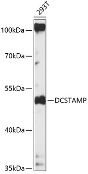
WB analysis of 293T cell lysate using DCSTAMP antibody. Dilution : 1:1000 Loading : 25μg per laneICC/IF
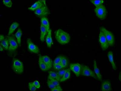
Immunofluorescence staining of HepG2 cells with DC-STAMP Antibody at 1:50, counter-stained with DAPI. The cells were fixed in 4% formaldehyde, permeabilized using 0.2% Triton X-100 and blocked in 10% normal Goat Serum. The cells were then incubated with the antibody overnight at 4°C. The secondary antibody was Alexa Fluor 488-congugated Goat Anti-Rabbit IgG(H+L).WB
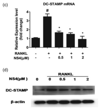
Total proteins were extracted and analysed by western blotting after RANKL stimulation for 48 h using antibodies against DC-STAMP and β-actin (c, d).WB
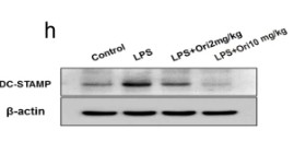
The BMMs from the left tibias and femurs of mice in each group were collected to measure the mRNA expression of f TRAP and g OSCAR and h ,the protein expression of DC-STAMP.WB
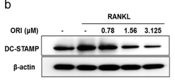
RAW264.7 cells were pretreated with or without ORI for 2 h and then stimulated with RANKL (100 ng/mL) for 48 h; b DC-STAMP protein levels were determined by Western blotting.WB
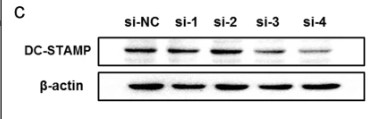
We synthesized four target siRNA sequences and verified their knockdown efficiency by c WB; the siRNA with the highest knockout efficiency was used for the follow-up experiments.WB
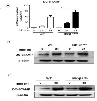
(A) DC-STAMP mRNA was detected by real-time PCR, (B) (C) DC-STAMP protein expression was measured by western blot.| Product Name | DC-STAMP Antibody (M-91) |
|---|---|
| Antibody Type | Primary Antibodies |
| Immunogen | epitope corresponding to amino acids 119-209 mapping within an internal region of DC-STAMP of mouse origin |
| Modification Notes | Bone homeostasis is accomplished by the balance of osteoblast and osteoclast activity. DC-STAMP (DC-specific transmembrane protein), also known as Transmembrane 7 superfamily member 4, is a 470 amino acid multi-pass membrane protein that regulates osteoclast and macrophage cell fusion. With localization at the cell surface, DC-STAMP is highly expressed in dendritic cells of the lung, kidney, liver and lymph nodes and is expressed at lower levels in spleen, bone marrow, leukocytes and pancreas. Mice that lack the gene encoding DC-STAMP exhibit upregulation of bone formation by osteoblasts and decreased bone reabsorption, ulitmately leading to an increase in bone mass. Alternatively, mice that overexpress the DC-STAMP gene show decreased osteoblast activity and bone mass, due to accelerated cell-cell fusion of osteoclasts. This evidence suggests that DC-STAMP plays an essential role in osteoclastogenesis. |
| Clonality | Polyclonal |
|---|---|
| Isotype | IgG |
| Host Species | Rabbit |
| Tested Applications | ELISAIFIPWB |
| WB:1:1000 ICC/IF:1:50 |
|
| Species Reactivity | HumanMouseRat |
| Concentration | 1mg/ml |
| Purification | Affinity purified |
| Gene Symbol | DCSTAMP |
|---|---|
| Gene Synonyms | FIND TM7SF4 hDC-STAMP |
| Gene Full Name | dendrocyte expressed seven transmembrane protein |
| Tissue Specificity | epitope corresponding to amino acids 119-209 mapping within an internal region of DC-STAMP of mouse origin |
| Entrez Gene | 81501 |
|---|
WB
WB analysis of 293T cell lysate using DCSTAMP antibody. Dilution : 1:1000 Loading : 25μg per lane
ICC/IF
Immunofluorescence staining of HepG2 cells with DC-STAMP Antibody at 1:50, counter-stained with DAPI. The cells were fixed in 4% formaldehyde, permeabilized using 0.2% Triton X-100 and blocked in 10% normal Goat Serum. The cells were then incubated with the antibody overnight at 4°C. The secondary antibody was Alexa Fluor 488-congugated Goat Anti-Rabbit IgG(H+L).
WB
Total proteins were extracted and analysed by western blotting after RANKL stimulation for 48 h using antibodies against DC-STAMP and β-actin (c, d).
WB
The BMMs from the left tibias and femurs of mice in each group were collected to measure the mRNA expression of f TRAP and g OSCAR and h ,the protein expression of DC-STAMP.
WB
RAW264.7 cells were pretreated with or without ORI for 2 h and then stimulated with RANKL (100 ng/mL) for 48 h; b DC-STAMP protein levels were determined by Western blotting.
WB
We synthesized four target siRNA sequences and verified their knockdown efficiency by c WB; the siRNA with the highest knockout efficiency was used for the follow-up experiments.
WB
(A) DC-STAMP mRNA was detected by real-time PCR, (B) (C) DC-STAMP protein expression was measured by western blot.| Application Notes | WB:1:1000 ICC/IF:1:50 |
|---|
| Form | Liquid |
|---|---|
| Storage Instructions | For short-term storage, store at 4° C. For long-term storage, aliquot and store at -20ºC or below. Avoid multiple freeze-thaw cycles. |
| Storage Buffer | phosphate buffered saline , pH 7.4, 150mM NaCl, 0.02% sodium azide and 50% glycerol. |


