Application
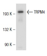
Western blot analysis of TRPM4 expression in MDCK whole cell lysate.IF
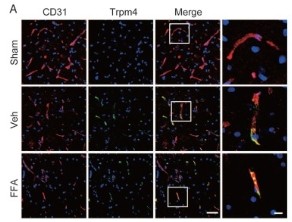
(A) Double immunofluorescence labeling of capillaries for Trpm4 (green) and CD31 (red) in sections through the penumbra at 1 d after injury in tissues obtained from sham-operated, vehicle control and FFA-treated mice. There was almost no obvious Trpm4 expression in the capillaries (labeled with CD31) of the sham-operated mice. As shown in the vehicle control and FFA-treated mice, Trpm4 was prominently up-regulated in the capillaries. Scale bar = 50 μm (left images) and 15 μm (right, higher magnification images, as marked in a white box in the left images).WB
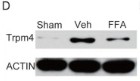
(D) Western blots performed for the Trpm4 protein in tissues obtained at 1 d after SCI. β-actin was used as the control.| Product Name | TRPM4 Antibody (H-55) |
|---|---|
| Antibody Type | Primary Antibodies |
| Modification Notes | Transient receptor potential ion channels (TRPCs) are a superfamily of six transmembrane segment-spanning, gated cation channels. TRPC subtypes mediate store-operated Ca2+ entry, a process involving Ca2+ influx and replenishment of Ca2+ stores formerly emptied through the action of inositol 1,4,5-trisphospate production and other Ca2+ mobilizing agents. TRP ion channels influence calcium-depletion induced calcium influx processes in response to chemo-, mechano- and osmoregulatory events. TRPM4 is a transient receptor potential channel with an intrinsic voltage-sensing mechanism. Voltage dependence of TRPM4 may be functionally important, especially in excitable tissues generating plateau-like or bursting action potentials. TRPM4-mediated depolarization modulates Ca2+ oscillations, with downstream effects on cytokine production in T lymphocytes. |
| Clonality | Polyclonal |
|---|---|
| Isotype | IgG |
| Host Species | Rabbit |
| Tested Applications | ELISAIFIPWB |
| recommended for detection of TRPM4 of human origin by WB, IP, IF and ELISA also reactive with additional species, including canine and bovineWB:1:100~1:200 IF:1:20~1:100 ELISA:1:1000~1:5000 IP:1:20~1:100 |
|
| Species Reactivity | HumanMouseRat |
| Concentration | 1mg/ml |
| Purification | Affinity purified |
| Alternative Names | epitope corresponding to amino acids 306-360 mapping within an N-terminal cytoplasmic domain of TRMP4 of human origin |
|---|---|
| Tissue Specificity | epitope corresponding to amino acids 306-360 mapping within an N-terminal cytoplasmic domain of TRMP4 of human origin |
| Entrez Gene | 54795 |
|---|
Application
Western blot analysis of TRPM4 expression in MDCK whole cell lysate.
IF
(A) Double immunofluorescence labeling of capillaries for Trpm4 (green) and CD31 (red) in sections through the penumbra at 1 d after injury in tissues obtained from sham-operated, vehicle control and FFA-treated mice. There was almost no obvious Trpm4 expression in the capillaries (labeled with CD31) of the sham-operated mice. As shown in the vehicle control and FFA-treated mice, Trpm4 was prominently up-regulated in the capillaries. Scale bar = 50 μm (left images) and 15 μm (right, higher magnification images, as marked in a white box in the left images).
WB
(D) Western blots performed for the Trpm4 protein in tissues obtained at 1 d after SCI. β-actin was used as the control.| Application Notes | recommended for detection of TRPM4 of human origin by WB, IP, IF and ELISA also reactive with additional species, including canine and bovineWB:1:100~1:200 IF:1:20~1:100 ELISA:1:1000~1:5000 IP:1:20~1:100 |
|---|
| Form | Liquid |
|---|---|
| Storage Instructions | Store at 4°C short term (1-2 weeks). Upon delivery aliquot. Store at -20°C. Avoid freeze / thaw cycle.Stable for 12 months from date of receipt |
| Storage Buffer | phosphate buffered saline , pH 7.4, 150mM NaCl, 0.02% sodium azide and 50% glycerol.Store at -20 °C. |


