Application
Immunoperoxidase staining of formalin fixed, paraffin-embedded human upper stomach tissue showing cytoplasmic staining of glandular cells.WB
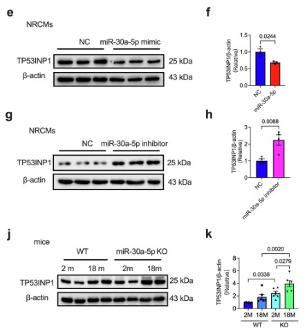
epresentative western blotting images (e) and quantitative analysis (f) of TP53INP1 expression in NRCMs transfected with NC or miR-30a-5p mimics (n = 3 in each group). Representative western blotting (g) and quantitative analysis (h) of TP53INP1 expression in NRCMs transfected with NC or the miR-30a-5p inhibitor (n = 4 in each group). i Luciferase activity assay of NRCMs cotransfected with NC or miR-30a-5p mimics and WT or mutant TP53INP1 (n = 5 in each group). Representative western blots (j) and quantitative analysis (k) of TP53INP1 expression in the hearts of 2- or 18-month-old WT or KO mice (n = 6 in each group).WB
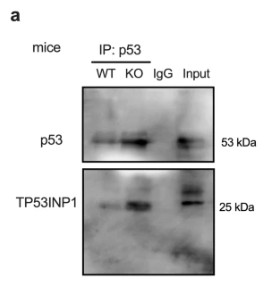
a Representative immunoprecipitation detection of the interaction between p53 and TP53INP1 in the heart tissues of WT and KO mice.h Representative immunoprecipitation blot showing the interaction of TP53INP1 with SUMO1 and p53 in the hearts of WT and KO mice.WB
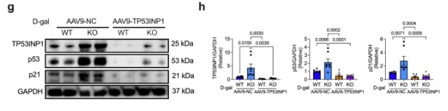
Representative western blotting images (g) and analysis (h) of TP53INP1 (n = 7 in each group), p53 (n = 7 in each group), and p21 (n = 7 in each group) expression in heart tissues of AAV9-cTnT-control- or AAV9-cTnT-TP53INP1-RNAi-transfected WT or KO mice induced with D-gal.| Product Name | TP53INP1 Antibody (H-110) |
|---|---|
| Antibody Type | Primary Antibodies |
| Modification Notes | TP53INP1 (tumor protein p53-inducible nuclear protein 1), also known as p53DINP1, SIP or Teap, is a 240 amino acid protein that localizes to nuclear bodies and exists as two alternatively spliced isoforms, designated p53DINP1a and p53DINP1b. Expressed ubiquitously with higher expression in testis, pancreas and spleen tissue, TP53INP1 functions in response to double-stranded DNA breaks and regulates p53-mediated apoptosis, specifically by phosphorylating human p53 at Ser 46, an event that leads to cell death. Additionally, TP53INP1 is thought to interact with p73 and may be involved in the regulation of p73-controlled cell cycle progression. TP53INP1 expression is downregulated in pancreatic ductal adenocarcinomas, suggesting that, via its ability to induce cell death, TP53INP1 plays a role in tumor suppression. |
| Clonality | Polyclonal |
|---|---|
| Isotype | IgG |
| Host Species | Rabbit |
| Tested Applications | ELISAIFIPWB |
| recommended for detection of TP53INP1 of mouse, human and, to a lesser extent, rat origin by WB, IP, IF and ELISA also reactive with additional species, including equine, canine, bovine and porcine | |
| Species Reactivity | HumanMouseRat |
| Concentration | 1mg/ml |
| Purification | Affinity purified |
| Alternative Names | epitope corresponding to amino acids 131-240 mapping at the C-terminus of TP53INP1 of human origin |
|---|---|
| Tissue Specificity | epitope corresponding to amino acids 131-240 mapping at the C-terminus of TP53INP1 of human origin |
| Entrez Gene | 94241 |
|---|
Application
Immunoperoxidase staining of formalin fixed, paraffin-embedded human upper stomach tissue showing cytoplasmic staining of glandular cells.
WB
epresentative western blotting images (e) and quantitative analysis (f) of TP53INP1 expression in NRCMs transfected with NC or miR-30a-5p mimics (n = 3 in each group). Representative western blotting (g) and quantitative analysis (h) of TP53INP1 expression in NRCMs transfected with NC or the miR-30a-5p inhibitor (n = 4 in each group). i Luciferase activity assay of NRCMs cotransfected with NC or miR-30a-5p mimics and WT or mutant TP53INP1 (n = 5 in each group). Representative western blots (j) and quantitative analysis (k) of TP53INP1 expression in the hearts of 2- or 18-month-old WT or KO mice (n = 6 in each group).
WB
a Representative immunoprecipitation detection of the interaction between p53 and TP53INP1 in the heart tissues of WT and KO mice.h Representative immunoprecipitation blot showing the interaction of TP53INP1 with SUMO1 and p53 in the hearts of WT and KO mice.
WB
Representative western blotting images (g) and analysis (h) of TP53INP1 (n = 7 in each group), p53 (n = 7 in each group), and p21 (n = 7 in each group) expression in heart tissues of AAV9-cTnT-control- or AAV9-cTnT-TP53INP1-RNAi-transfected WT or KO mice induced with D-gal.| Application Notes | recommended for detection of TP53INP1 of mouse, human and, to a lesser extent, rat origin by WB, IP, IF and ELISA also reactive with additional species, including equine, canine, bovine and porcine |
|---|
| Form | Liquid |
|---|---|
| Storage Instructions | For short-term storage, store at 4° C. For long-term storage, aliquot and store at -20ºC or below. Avoid multiple freeze-thaw cycles. |
| Storage Buffer | phosphate buffered saline , pH 7.4, 150mM NaCl, 0.02% sodium azide and 50% glycerol. |


