Application
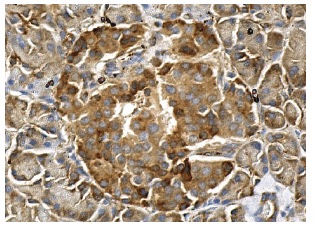
Immunoperoxidase staining of formalin fixed, paraffin-embedded human pancreas tissue showing cytoplasmic staining of Islets of Langerhans and glandular cells.Application
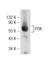
Western blot analysis of PDK expression in mouse heart tissue extract.Application
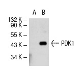
Western blot analysis of PDK1 expression in non-transfected: (A) and human PDK1 transfected: (B) 293T whole cell lysates.Application
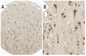
Immunoperoxidase staining of formalin fixed, paraffin-embedded human cerebral cortex tissue showing cytoplasmic staining of neuronal and glial cells (low and high magnification). Kindly provided by The Swedish Human Protein Atlas (HPA) program.Application
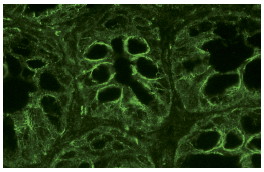
Immunofluorescence staining of normal mouse intestine frozen section showing cytoplasmic staining.| Product Name | PDK Antibody (H-300) |
|---|---|
| Antibody Type | Primary Antibodies |
| Modification Notes | Pyruvate dehydrogenase kinase family members (PDK1, 2, 3 and 4) are Serine kinases that catalyze phosphorylation of the E1å subunit of the pyruvate dehydrogenase complex (PDC). PDC activity is controlled through phosphoryl-ation and dephosphorylation of the E1å subunit, which leads to inactivation and reactivation, respectively. PDK3 binding to a free lipoyl domain (L2) in dihydrolypoyl acetyltransferase (E2), which comprises the core of PDC, leads to a large increase in E1å phosphorylation. Upregulation of PDK isoenzymes occurs during starvation conditions, where acetyl-CoA is alternatively generated through fatty acid oxidation. PDKs contain five conserved regions and are mechanistically similar to bacterial His-kinases, in that both require histidine residues for activity. In mammals, transcripts for PDK3 are most abundant in testis and are moderately expressed in heart and skeletal muscle. |
| Clonality | Polyclonal |
|---|---|
| Isotype | IgG |
| Host Species | Rabbit |
| Tested Applications | WBIPIFIHCELISA |
| recommended for detection of PDK1, 2, 3 and 4 of mouse, rat and human origin by WB, IP, IF, IHC(P) and ELISA also reactive with additional species, including equine, canine, bovine, porcine and avian | |
| Species Reactivity | HumanMouseRat |
| Concentration | 1mg/ml |
| Purification | Affinity purified |
| Alternative Names | epitope corresponding to amino acids 137-436 mapping at the C-terminus of PDK1 of human origin |
|---|---|
| Tissue Specificity | epitope corresponding to amino acids 137-436 mapping at the C-terminus of PDK1 of human origin |
| Entrez Gene | 5163 |
|---|
Application
Immunoperoxidase staining of formalin fixed, paraffin-embedded human pancreas tissue showing cytoplasmic staining of Islets of Langerhans and glandular cells.
Application
Western blot analysis of PDK expression in mouse heart tissue extract.
Application
Western blot analysis of PDK1 expression in non-transfected: (A) and human PDK1 transfected: (B) 293T whole cell lysates.
Application
Immunoperoxidase staining of formalin fixed, paraffin-embedded human cerebral cortex tissue showing cytoplasmic staining of neuronal and glial cells (low and high magnification). Kindly provided by The Swedish Human Protein Atlas (HPA) program.
Application
Immunofluorescence staining of normal mouse intestine frozen section showing cytoplasmic staining.| Application Notes | recommended for detection of PDK1, 2, 3 and 4 of mouse, rat and human origin by WB, IP, IF, IHC(P) and ELISA also reactive with additional species, including equine, canine, bovine, porcine and avian |
|---|
| Form | Liquid |
|---|---|
| Storage Instructions | For short-term storage, store at 4° C. For long-term storage, aliquot and store at -20ºC or below. Avoid multiple freeze-thaw cycles. |
| Storage Buffer | phosphate buffered saline , pH 7.4, 150mM NaCl, 0.02% sodium azide and 50% glycerol. |


