Application
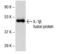
Western blot analysis of human recombinant IL-1β fusion protein.Application
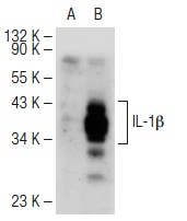
Western blot analysis of IL-1β expression in non-transfected: (A) and human IL-1β transfected: (B) 293T whole cell lysates.Application
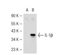
Western blot analysis of IL-1β expression in non-transfected: (A) and human IL-1β transfected: (B) 293T whole cell lysates.Application
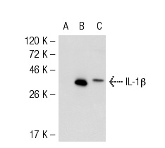
Western blot analysis of IL-1β expression in non-transfected 293: (A), human IIL-1β transfected 293: (B) and BJAB (C) whole cell lysates.Application
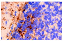
Immunoperoxidase staining of formalin fixed, paraffin-embedded mouse brain tissue showing extracellular localization.WB
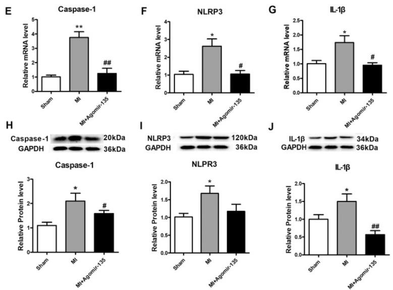
(E-J) The expression of caspase-1, NLRP3 and IL-1β both mRNA levels and protein levels were increased after MI-1 day compared to sham group and were inhibited in agomir-135b group compared to MI group.WB
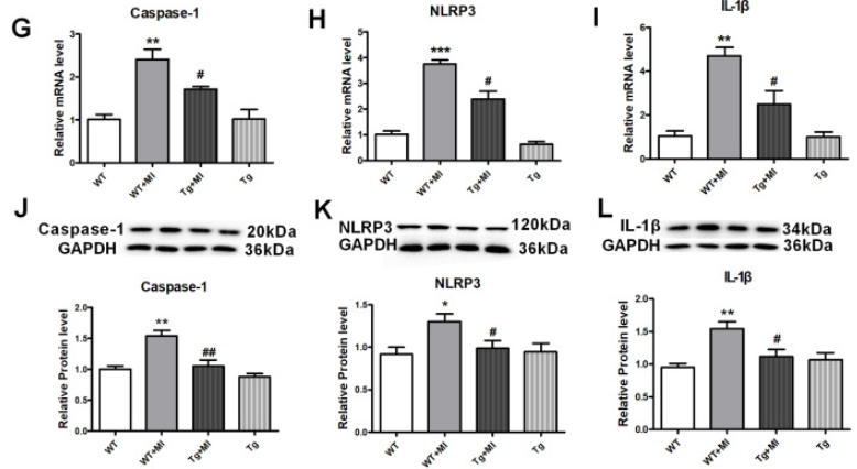
(G–L) The expression of caspase-1, NLRP3 and IL-1β both mRNA levels and protein levels were increased after MI-1 day compared to WT and were inhibited in overexpressing miR-135b Tg mice.WB
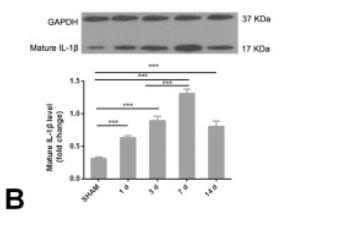
B: The levels of the mature IL-1β protein in mouse retinas from the surgery and the sham surgery groups were analyzed at different time points after OPC injury in the sterile environment using Western blotting.IF
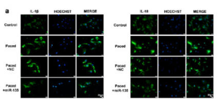
a Representative image of immunofluorescence for IL-1β and IL-18, scale bars = 20 μm.IF
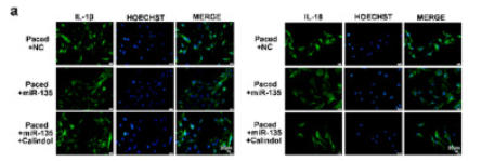
a Representative image of immunofluorescence for IL-1β and IL-18, scale bars = 20 μm.IHC
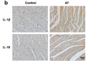
b Immunohistochemistry of IL-1β and IL-18; scale bars = 20 μm.IHC
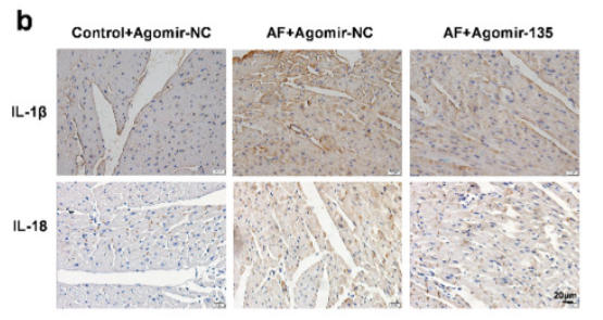
b Immunohistochemistry of IL-1β and IL-18, scale bars = 20 μm.| Product Name | IL-1β Antibody (H-153) |
|---|---|
| Antibody Type | Primary Antibodies |
| Product description |
|
| Modification Notes | Two forms of interleukin-1, designated IL-1å and IL-1∫, have been described. Although encoded by distinct genes and exhibiting roughly only 25% sequence identity, IL-1å and IL-1∫ bind to the same receptor and seem to elicit similar biological responses. IL-1 production is generally thought to be associated with inflammation, but it has also been shown to be expressed during kidney development, thymocyte differentiation and cartilage degradation. IL-1 plays a critical role in the regulation of immune response and inflammation, acting as an activator of T and B lymphocytes and natural killer (NK) cells. In T cells, IL-1 stimulates the production of IL-2 and selectively inhibits IL-4 expression. IL-1 induces B cell proliferation and maturation, and immunoglobulin synthesis. NK cells require IL-1∫ for production of the anti-pathogen IFN-©. IL-1 has also been implicated in several pathological conditions including rheumatoid arthritis, inflammatory bowel disease and atherosclerosis. |
| Isotype | IgG |
|---|---|
| Host Species | Rabbit |
| Tested Applications | ELISAIFIHCIPWB |
| recommended for detection of IL-1β of mouse, rat and human origin by WB, IP, IF, IHC(P) and ELISA: WB:1:100~1:200 IP:1:20~1:100 IF:1:20~1:100 IHC:1:10~1:50 ELISA:1:1000~1:5000 |
|
| Species Reactivity | HumanMouseRat |
| Concentration | 1mg/ml |
| Purification | Affinity purified |
| Alternative Names | epitope corresponding to amino acids 117-269 mapping at the C-terminus of IL-1&beta of human origin |
|---|---|
| Tissue Specificity | epitope corresponding to amino acids 117-269 mapping at the C-terminus of IL-1β of human origin |
| Entrez Gene | 3553 |
|---|
Application
Western blot analysis of human recombinant IL-1β fusion protein.
Application
Western blot analysis of IL-1β expression in non-transfected: (A) and human IL-1β transfected: (B) 293T whole cell lysates.
Application
Western blot analysis of IL-1β expression in non-transfected: (A) and human IL-1β transfected: (B) 293T whole cell lysates.
Application
Western blot analysis of IL-1β expression in non-transfected 293: (A), human IIL-1β transfected 293: (B) and BJAB (C) whole cell lysates.
Application
Immunoperoxidase staining of formalin fixed, paraffin-embedded mouse brain tissue showing extracellular localization.
WB
(E-J) The expression of caspase-1, NLRP3 and IL-1β both mRNA levels and protein levels were increased after MI-1 day compared to sham group and were inhibited in agomir-135b group compared to MI group.
WB
(G–L) The expression of caspase-1, NLRP3 and IL-1β both mRNA levels and protein levels were increased after MI-1 day compared to WT and were inhibited in overexpressing miR-135b Tg mice.
WB
B: The levels of the mature IL-1β protein in mouse retinas from the surgery and the sham surgery groups were analyzed at different time points after OPC injury in the sterile environment using Western blotting.
IF
a Representative image of immunofluorescence for IL-1β and IL-18, scale bars = 20 μm.
IF
a Representative image of immunofluorescence for IL-1β and IL-18, scale bars = 20 μm.
IHC
b Immunohistochemistry of IL-1β and IL-18; scale bars = 20 μm.
IHC
b Immunohistochemistry of IL-1β and IL-18, scale bars = 20 μm.| Application Notes | recommended for detection of IL-1β of mouse, rat and human origin by WB, IP, IF, IHC(P) and ELISA: WB:1:100~1:200 IP:1:20~1:100 IF:1:20~1:100 IHC:1:10~1:50 ELISA:1:1000~1:5000 |
|---|
| Form | Liquid |
|---|---|
| Storage Instructions | For short-term storage, store at 4° C. For long-term storage, aliquot and store at -20ºC or below. Avoid multiple freeze-thaw cycles. |
| Storage Buffer | phosphate buffered saline , pH 7.4, 150mM NaCl, 0.02% sodium azide and 50% glycerol. |


