Application
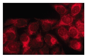
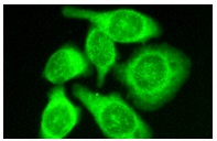
Immunofluorescence staining of methanol-fixed HeLa cells showing cytoplasmic localization.Application
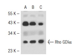
Western blot analysis of Rho GDIα expression in non-transfected 293T: (A), human Rho GDIα transfected 293T: (B) and MCF7 (C) whole cell lysates.Application
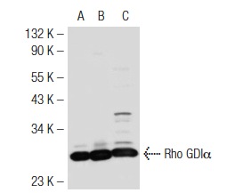
Western blot analysis of Rho GDIα expression in non-transfected 293T: (A), mouse Rho GDIα transfected 293T: (B) and K-562 (C) whole cell lysates.Application
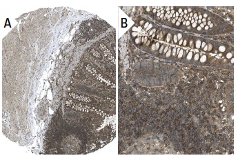
Immunoperoxidase staining of formalin fixed, paraffin-embedded human appendix tissue showing cytoplasmic staining of lymphoid and glandular cells at low (A) and high (B) magnification. Kindly provided by The Swedish Human Protein Atlas (HPA) program.Application
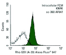
Intracellular FCM analysis of fixed and permeabilized KNRK cells. Black line histogram represents the isotype control, normal rabbit IgG: .Application
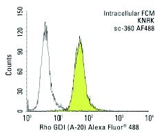
Intracellular FCM analysis of fixed and permeabilized KNRK cells. Black line histogram represents the isotype control, normal rabbit IgG: .Application
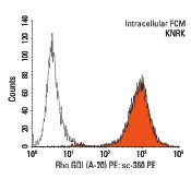
Intracellular FCM analysis of fixed and permeabilized KNRK cells. Black line histogram represents the isotype control, normal rabbit IgG: .Application
Immunofluorescence staining of methanol-fixed HeLa cells showing cytoplasmic staining.Application
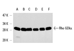
Western blot analysis of Rho GDIα expression in KNRK (A), PC-12 (B), HL-60 (C), HeLa (D), SK-BR-3 (E) and MCF7 (F) whole cell lysates.| Product Name | Rho GDIα Antibody (A-20) |
|---|---|
| Antibody Type | Primary Antibodies |
| Antigen Alias | epitope mapping at the N-terminus of Rho GDIα of human origin |
| Modification Notes | Members of the Ras superfamily of small GTP-binding proteins are critical mediators of diverse cell signaling pathways, including those leading to cell proliferation, cytoskeletal organization and secretion. The counter-conversion of the active GTP-bound form of these proteins to their inactive GDP-bound form is influenced by two types of regulatory proteins: those that alter the intrinsic GTPase activity of the GTP-binding proteins and those that alter the rate of GDP/GTP exchange. Guanine nucleotide-releasing factors (GRFs) increase the GDP dissociation rate, while GDP-dissociation inhibitors (GDIs) decrease the dissociation rate. Rho GDIå, also known as ARHGDIA or GDIA1, is a 204 amino acid member of the Rho GDI family of proteins. Localized to the cytoplasm, Rho GDIå inhibits the dissociation of GDP from Rho proteins, thereby preventing GTP from binding to and subsequently activating Rho proteins. In humans, Rho GDIå can be phosphorylated at Ser 101 by p21-activated kinase (åPAK), an event that inhibits Rho GDIå activity and may result in positive feedback regulation of certain Rho GDIå target proteins. |
| Clonality | Polyclonal |
|---|---|
| Isotype | IgG |
| Host Species | Rabbit |
| Tested Applications | WBIPIFIHCFCELISA |
| recommended for detection of Rho GDIα of mouse, rat and human origin by WB, IP, IF, IHC(P), FCM and ELISA also reactive with additional species, including equine, canine, bovine, porcine and avian: |
|
| Species Reactivity | HumanMouseRat |
| Concentration | 1mg/ml |
| Purification | Affinity purified |
| Alternative Names | epitope mapping at the N-terminus of Rho GDI&alpha of human origin |
|---|---|
| Tissue Specificity | epitope mapping at the N-terminus of Rho GDIα of human origin |
| Entrez Gene | 396 |
|---|
Application
Immunofluorescence staining of methanol-fixed HeLa cells showing cytoplasmic localization.
Application
Western blot analysis of Rho GDIα expression in non-transfected 293T: (A), human Rho GDIα transfected 293T: (B) and MCF7 (C) whole cell lysates.
Application
Western blot analysis of Rho GDIα expression in non-transfected 293T: (A), mouse Rho GDIα transfected 293T: (B) and K-562 (C) whole cell lysates.
Application
Immunoperoxidase staining of formalin fixed, paraffin-embedded human appendix tissue showing cytoplasmic staining of lymphoid and glandular cells at low (A) and high (B) magnification. Kindly provided by The Swedish Human Protein Atlas (HPA) program.
Application
Intracellular FCM analysis of fixed and permeabilized KNRK cells. Black line histogram represents the isotype control, normal rabbit IgG: .
Application
Intracellular FCM analysis of fixed and permeabilized KNRK cells. Black line histogram represents the isotype control, normal rabbit IgG: .
Application
Intracellular FCM analysis of fixed and permeabilized KNRK cells. Black line histogram represents the isotype control, normal rabbit IgG: .
Application
Immunofluorescence staining of methanol-fixed HeLa cells showing cytoplasmic staining.
Application
Western blot analysis of Rho GDIα expression in KNRK (A), PC-12 (B), HL-60 (C), HeLa (D), SK-BR-3 (E) and MCF7 (F) whole cell lysates.| Application Notes | recommended for detection of Rho GDIα of mouse, rat and human origin by WB, IP, IF, IHC(P), FCM and ELISA also reactive with additional species, including equine, canine, bovine, porcine and avian: |
|---|
| Form | Liquid |
|---|---|
| Storage Instructions | For short-term storage, store at 4° C. For long-term storage, aliquot and store at -20ºC or below. Avoid multiple freeze-thaw cycles. |
| Storage Buffer | phosphate buffered saline , pH 7.4, 150mM NaCl, 0.02% sodium azide and 50% glycerol. |


