Application
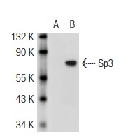
Western blot analysis of Sp3 expression in non-transfected: (A) and mouse Sp3 transfected: (B) 293T whole cell lysates.Application
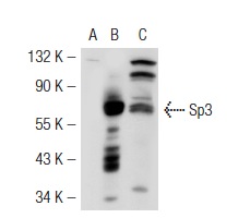
Western blot analysis of Sp3 expression in non-transfected: (A) and mouse Sp3 transfected: (B) 293T whole cell lysates and HeLa nuclear extract (C).Application
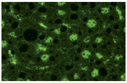
Immunofluorescence staining of normal mouse liver frozen section showing nuclear staining.Application
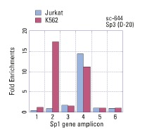
ChIP analysis of Sp3 recruitment to genomic amplicons. Four (1-4) different human genomic Sp-1 and two control amplicons (5,6) were analyzed by quantitative PCR (primer sequences available in on-line supplemental data). Data generated in collaboration with Drs. N. Trinklein and R. Myers, Stanford University (ENCODE Project).Application
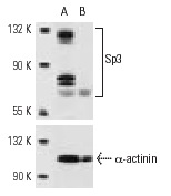
Western blot analysis of Sp3 expression in non-transfected control (A) and Sp3 siRNA transfected (B) HeLa cells. Blot probed with Sp3 (D-20): . α-actinin (H-2): used as specificity and loading control.Application
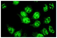
Nuclear immunofluorescence staining of methanol-fixed HeLa cells.Application
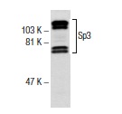
Western blot analysis of Sp3 expression in KNRK nuclear extract.| Product Name | Sp3 Antibody (D-20) FITC |
|---|---|
| Antibody Type | Primary Antibodies |
| Antigen Alias | epitope mapping at the C-terminus of Sp3 of human origin |
| Modification Notes | The Sp transcription factor family includes Sp1, Sp2, Sp3 (SPR-2) and Sp4 (SPR-1). Sp transcription factors share similar structures but do not share simi-lar functions. All four proteins contain a highly conserved DNA-binding domain composed of three zinc fingers at the C-terminus. Sp family members bind the consensus sequence GGGGCGGGGC and other closely related sequences which are known as GC boxes. Sp1, Sp3 and Sp4 share a high affinity for GC boxes while Sp2 does not. Sp2 only weakly binds to GT boxes. Sp1, Sp2 and Sp3 are ubiquitously expressed, while Sp4 is abundantly expressed in brain with limited expression in other tissues. Sp1 and Sp3, but not Sp2 or Sp4, interact with E2, a regulatory element for the ∫4 subunit of neuronal nicotinic acetylcholine receptors. Sp3 is the only Sp member to inhibit Sp1 and Sp4 mediated transcription. Multiple isoforms of Sp3 exist due to alternative splicing events. |
| Clonality | Polyclonal |
|---|---|
| Isotype | IgG |
| Host Species | Rabbit |
| Tested Applications | WBIPIFELISAGSChIP |
| recommended for detection of Sp3 of mouse, rat and human origin by WB, IP, IF and ELISA also reactive with additional species, including equine, canine, bovine, porcine and avian: |
|
| Species Reactivity | HumanMouseRat |
| Concentration | 1mg/ml |
| Purification | Affinity purified |
| Alternative Names | epitope mapping at the C-terminus of Sp3 of human origin |
|---|---|
| Tissue Specificity | epitope mapping at the C-terminus of Sp3 of human origin |
| Entrez Gene | 6670 |
|---|
Application
Western blot analysis of Sp3 expression in non-transfected: (A) and mouse Sp3 transfected: (B) 293T whole cell lysates.
Application
Western blot analysis of Sp3 expression in non-transfected: (A) and mouse Sp3 transfected: (B) 293T whole cell lysates and HeLa nuclear extract (C).
Application
Immunofluorescence staining of normal mouse liver frozen section showing nuclear staining.
Application
ChIP analysis of Sp3 recruitment to genomic amplicons. Four (1-4) different human genomic Sp-1 and two control amplicons (5,6) were analyzed by quantitative PCR (primer sequences available in on-line supplemental data). Data generated in collaboration with Drs. N. Trinklein and R. Myers, Stanford University (ENCODE Project).
Application
Western blot analysis of Sp3 expression in non-transfected control (A) and Sp3 siRNA transfected (B) HeLa cells. Blot probed with Sp3 (D-20): . α-actinin (H-2): used as specificity and loading control.
Application
Nuclear immunofluorescence staining of methanol-fixed HeLa cells.
Application
Western blot analysis of Sp3 expression in KNRK nuclear extract.| Application Notes | recommended for detection of Sp3 of mouse, rat and human origin by WB, IP, IF and ELISA also reactive with additional species, including equine, canine, bovine, porcine and avian: |
|---|
| Form | Liquid |
|---|---|
| Storage Instructions | For short-term storage, store at 4° C. For long-term storage, aliquot and store at -20ºC or below. Avoid multiple freeze-thaw cycles. |
| Storage Buffer | phosphate buffered saline , pH 7.4, 150mM NaCl, 0.02% sodium azide and 50% glycerol. |


