Application
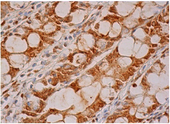
Immunoperoxidase staining of formalin fixed, paraffin-embedded human rectum tissue showing nuclear and cytoplasmic staining of glandular cells.Application
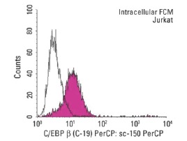
Indirect, intracellular FCM analysis of fixed and permeabilized Jurkat cells stained with C/EBP β (C-19),followed by PerCP-conjugated goat anti-rabbit IgG: . Black line histogram represents the isotype control, normal rabbit IgG: .Application
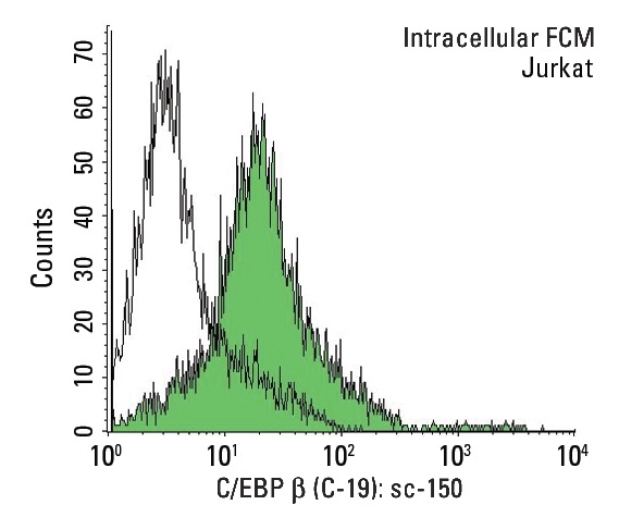
Indirect, intracellular FCM analysis of fixed and permeabilized Jurkat cells stained with C/EBP β (C-19), followed by FITC-conjugated bovine anti-rabbit IgG: . Black line histogram represents the isotype control, normal rabbit IgG: .Application
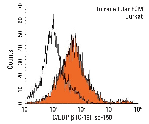
Indirect, intracellular FCM analysis of fixed and permeabilized Jurkat cells stained with C/EBP β (C-19), followed by PE-conjugated chicken anti-rabbit IgG: . Black line histogram represents the isotype control, normal rabbit IgG: .Application
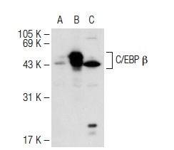
Western blot analysis of C/EBP β expression in non-transfected 293T: (A), human C/EBP β transfected 293T: (B) and Jurkat (C) whole cell lysates.Application
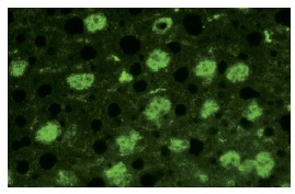
Immunofluorescence staining of normal mouse liver frozen section showing nuclear staining.Application
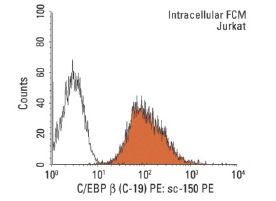
Indirect, intracellular FCM analysis of fixed and permeabilized Jurkat cells stained with C/EBP β (C-19), followed by PE-conjugated goat anti-rabbit IgG: . Black line histogram represents the isotype control, normal rabbit IgG: .Application
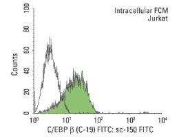
Indirect, intracellular FCM analysis of fixed and permeabilized Jurkat cells stained with C/EBP β (C-19), followed by FITC-conjugated goat anti-rabbit IgG: . Black line histogram represents the isotype control, normal rabbit IgG: .Application
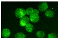
Immunofluorescence staining of methanol-fixed Jurkat cells showing nuclear staining.Application
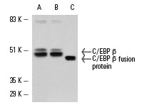
Western blot analysis of C/EBP β expression in control (A) and phorbol-induced (B) Jurkat cell nuclear extracts. C/EBP β fusion protein is shown as positive control (C).| Product Name | C/EBP β Antibody (C-19) AC |
|---|---|
| Antibody Type | Primary Antibodies |
| Antigen Alias | epitope mapping at the C-terminus of C/EBP β of rat origin |
| Modification Notes | CCAAT-enhancer binding proteins (C/EBP) are basic region/leucine zipper (bZIP) transcription factors selectively expressed during the differentiation of liver, adipose tissue, blood cells and the endocrine pancreas. C/EBP ∫ is a member of the C/EBP transcription factor family. The C/EBP ∫ gene encodes several isoforms containing truncated transcription activation domains due to the alternative translational initiation at multipe AUG start sites. C/EBP ∫ is also known as interleukin 6-dependent DNA-binding protein (IL6DBP), liver activator protein (LAP) or liver-enriched transcriptional activator protein transcription factor 5 (TCF5). C/EBP ∫ contributes to the regulation of the acute phase response in hepatocytes. Stat3 has an important function in IL-6-mediated transcription of the C/EBP ∫ gene that has direct implication for acute phase response in liver cells.The C/EBP ∫ form requires phosphorylation for its DNA binding ability, and increase binding of C/EBP ∫ isoforms during acute-phase reaction occurs through its upregulation and structural modification. |
| Clonality | Polyclonal |
|---|---|
| Isotype | IgG |
| Host Species | Rabbit |
| Tested Applications | WBIPIFFCELISAGSChIP |
| recommended for detection of C/EBP β of mouse, rat and human origin by WB, IP, IF, ELISA and indirect FCM using PE (sc-3739)- and FITC (sc-2012)-conjugated goat anti-rabbit IgG also reactive with additional species, including equine, bovine and porcine: |
|
| Species Reactivity | HumanMouseRat |
| Concentration | 1mg/ml |
| Purification | Affinity purified |
| Alternative Names | epitope mapping at the C-terminus of C/EBP &beta of rat origin |
|---|---|
| Tissue Specificity | epitope mapping at the C-terminus of C/EBP β of rat origin |
| Entrez Gene | 1051 |
|---|
Application
Immunoperoxidase staining of formalin fixed, paraffin-embedded human rectum tissue showing nuclear and cytoplasmic staining of glandular cells.
Application
Indirect, intracellular FCM analysis of fixed and permeabilized Jurkat cells stained with C/EBP β (C-19),followed by PerCP-conjugated goat anti-rabbit IgG: . Black line histogram represents the isotype control, normal rabbit IgG: .
Application
Indirect, intracellular FCM analysis of fixed and permeabilized Jurkat cells stained with C/EBP β (C-19), followed by FITC-conjugated bovine anti-rabbit IgG: . Black line histogram represents the isotype control, normal rabbit IgG: .
Application
Indirect, intracellular FCM analysis of fixed and permeabilized Jurkat cells stained with C/EBP β (C-19), followed by PE-conjugated chicken anti-rabbit IgG: . Black line histogram represents the isotype control, normal rabbit IgG: .
Application
Western blot analysis of C/EBP β expression in non-transfected 293T: (A), human C/EBP β transfected 293T: (B) and Jurkat (C) whole cell lysates.
Application
Immunofluorescence staining of normal mouse liver frozen section showing nuclear staining.
Application
Indirect, intracellular FCM analysis of fixed and permeabilized Jurkat cells stained with C/EBP β (C-19), followed by PE-conjugated goat anti-rabbit IgG: . Black line histogram represents the isotype control, normal rabbit IgG: .
Application
Indirect, intracellular FCM analysis of fixed and permeabilized Jurkat cells stained with C/EBP β (C-19), followed by FITC-conjugated goat anti-rabbit IgG: . Black line histogram represents the isotype control, normal rabbit IgG: .
Application
Immunofluorescence staining of methanol-fixed Jurkat cells showing nuclear staining.
Application
Western blot analysis of C/EBP β expression in control (A) and phorbol-induced (B) Jurkat cell nuclear extracts. C/EBP β fusion protein is shown as positive control (C).| Application Notes | recommended for detection of C/EBP β of mouse, rat and human origin by WB, IP, IF, ELISA and indirect FCM using PE (sc-3739)- and FITC (sc-2012)-conjugated goat anti-rabbit IgG also reactive with additional species, including equine, bovine and porcine: |
|---|
| Form | Liquid |
|---|---|
| Storage Instructions | For short-term storage, store at 4° C. For long-term storage, aliquot and store at -20ºC or below. Avoid multiple freeze-thaw cycles. |
| Storage Buffer | phosphate buffered saline , pH 7.4, 150mM NaCl, 0.02% sodium azide and 50% glycerol. |


