WB
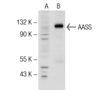
Western blot analysis of AASS expression in non-transfected: (A) and mouse AASS transfected: (B) 293T whole cell lysates.IHC
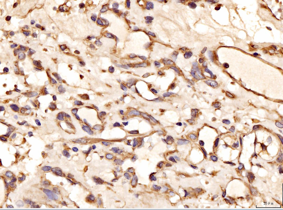
IHC analysis using anti-AASS Antibody. detected in paraffin-embedded section of human kidney cancer tissue. Biotinylated goat anti-rabbit IgG was used as secondary antibody. The tissue section was developed using Strepavidin Biotin-Complex (SABC) with DAB as the chromogen.ICC/IF
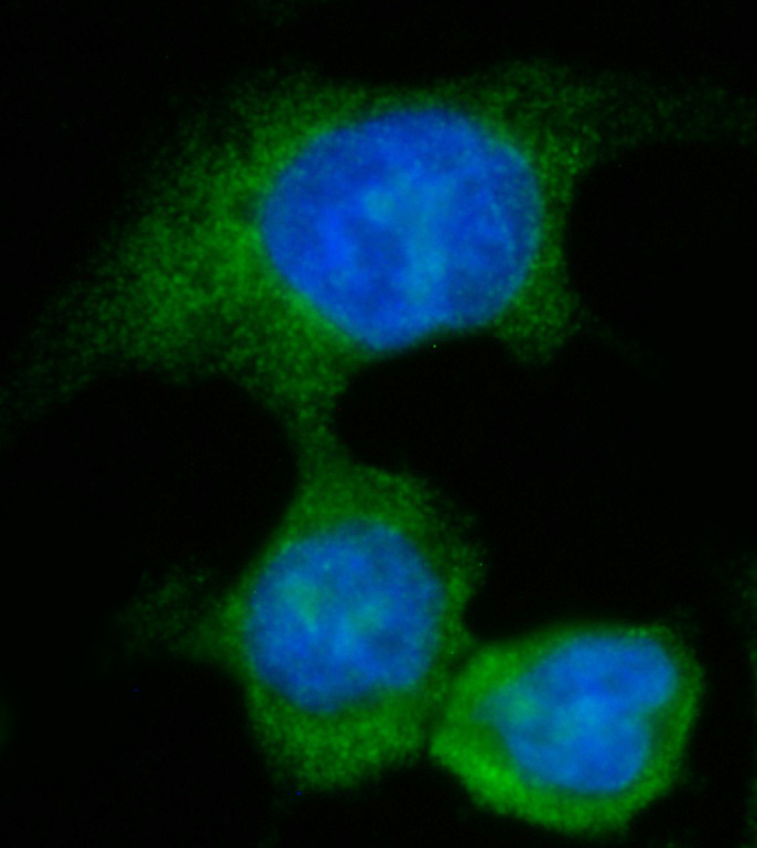
ICC analysis using anti- AASS Antibody. was detected in immersion fixed CACO-2 cell. Cells were stained using the Dylight488-conjugated Anti-rabbit IgG Secondary Antibody (green)and counterstained with DAPI (blue).| Product Name | AASS Antibody (H-300) |
|---|---|
| Antibody Type | Primary Antibodies |
| Antigen Alias | epitope corresponding to amino acids 627-926 mapping at the C-terminus of AASS of human origin |
| Conjugation | Agarose |
| Modification | Acetylated |
| Modification Notes | Alpha-aminoadipic semialdehyde synthase (AASS), also designated lysine ketoglutarate reductase (LKR) or saccharopine dehydrogenase (SDH), is a 926 amino acid protein that exists as a homodimer in the mitochondria. AASS acts as a bifunctional enzyme containing the lysine alpha-ketoglutarate reductase (LKR) and saccharopine dehydrogenase activities that catalyzes the first two steps in lysine degradation. It is widely expressed with highest expression in liver and transcription of the AASS gene is induced upon starvation. Mutations in the gene encoding AASS result in various forms familial hyperlysinemias (FH), autosomal recessive disorders characterized by hyperlysinemia, lysinuria, and variable saccharopinuria. However, no adverse mental or physical effects have been found in patients with hyperlysinemia. |
| Clonality | Polyclonal |
|---|---|
| Isotype | IgG |
| Host Species | Rabbit |
| Tested Applications | ELISAIFIHCWB |
| WB:1:500-2000 IHC:1:50-400 ICC:1:50-400 |
|
| Species Reactivity | HumanMouseRat |
| Concentration | 1mg/ml |
| Purification | Affinity purified |
| Alternative Names | epitope corresponding to amino acids 627-926 mapping at the C-terminus of AASS of human origin |
|---|---|
| Molecular Weight(MW) | 102KD |
| Tissue Specificity | epitope corresponding to amino acids 627-926 mapping at the C-terminus of AASS of human origin |
| Entrez Gene | 10157 |
|---|
WB
Western blot analysis of AASS expression in non-transfected: (A) and mouse AASS transfected: (B) 293T whole cell lysates.
IHC
IHC analysis using anti-AASS Antibody. detected in paraffin-embedded section of human kidney cancer tissue. Biotinylated goat anti-rabbit IgG was used as secondary antibody. The tissue section was developed using Strepavidin Biotin-Complex (SABC) with DAB as the chromogen.
ICC/IF
ICC analysis using anti- AASS Antibody. was detected in immersion fixed CACO-2 cell. Cells were stained using the Dylight488-conjugated Anti-rabbit IgG Secondary Antibody (green)and counterstained with DAPI (blue).| Application Notes | WB:1:500-2000 IHC:1:50-400 ICC:1:50-400 |
|---|
| Form | Liquid |
|---|---|
| Storage Instructions | For short-term storage, store at 4° C. For long-term storage, aliquot and store at -20ºC or below. Avoid multiple freeze-thaw cycles. |
| Storage Buffer | phosphate buffered saline , pH 7.4, 150mM NaCl, 0.02% sodium azide and 50% glycerol. |


