Application
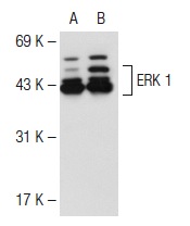
Western blot analysis of ERK 1 expression in non-transfected: (A) and human ERK 1 transfected: (B) 293T whole cell lysates.Application
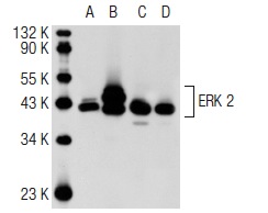
Western blot analysis of ERK 2 expression in non-transfected 293T: (A), human ERK 2 transfected 293T: (B), A-431 (C) and HeLa (D) whole cell lysates.Application
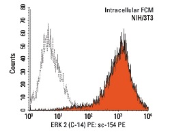
Intracellular FCM analysis of fixed and permeabilized NIH/3T3 cells. Black line histogram represents the isotype control, normal rabbit IgG: .Application
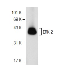
Western blot analysis of ERK 2 expression in rat hippocampus tissue extract.Application
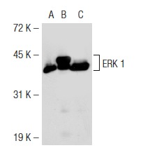
Western blot analysis of ERK 1 expression in non-transfected 293T: (A), mouse ERK 1 transfected 293T: (B) and A-431 (C) whole cell lysates.Application
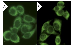
Immunofluorescence staining of methanol-fixed HeLa cells showing cytoplasmic localization using indirect FITC (A) staining and direct Alexa Fluor 488 (B) staining.Application
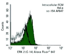
Intracellular FCM analysis of fixed and permeabilized HeLa cells. Black line histogram represents the isotype control, normal rabbit IgG: .Application
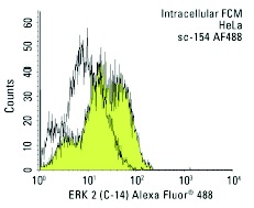
Intracellular FCM analysis of fixed and permeabilized HeLa cells. Black line histogram represents the isotype control, normal rabbit IgG: .Application
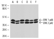
Antibodies tested include ERK 1 (K-23)-G: (A-C) and ERK 2 (C-14)-G: (D-F).Application
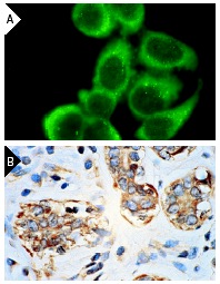
Immunofluorescence staining of methanol-fixed HeLa cells (A) and immunoperoxidase staining of formalin-fixed, paraffin-embedded human breast carcinoma tissue (B) showing cytoplasmic localization.Application
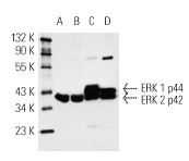
Western blot analysis of ERK 1 and ERK 2 expression in A-431 (A), HeLa (B), KNRK (C) and NIH/3T3 (D) whole cell lysates.Application
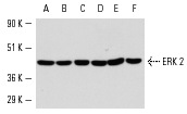
Western blot analysis of ERK 2 expression in A-431 (A), HeLa (B), KNRK (C), NIH/3T3 (D), NRK (E) and DU 145 (F) whole cell lysates.| Product Name | ERK 2 Antibody (C-14) AC |
|---|---|
| Antibody Type | Primary Antibodies |
| Antigen Alias | epitope mapping at the C-terminus of ERK 2 of rat origin |
| Modification Notes | Mitogen-activated protein kinase (MAPK) signaling pathways involve two closely related MAP kinases, known as extracellular-signal-related kinase 1 (ERK 1, p44) and 2 (ERK 2, p42). Growth factors, steroid hormones, G protein-coupled receptor ligands, and neurotransmitters can initiate MAPK signaling pathways. Activation of ERK1 and ERK2 requires phosphorylation by upstream kinases such as MAP kinase kinase (MEK), MEK kinase and Raf-1. ERK1 and ERK2 phosphorylation can occur at specific tyrosine and threonine sites mapping within consensus motifs that include the Threonine-Glutamate-Tyrosine motif. ERK activation leads to dimerization with other ERKs and subsequent localization to the nucleus. Active ERK dimers phosphorylate serine and threonine residues on nuclear proteins and influence a host of responses that include proliferation, differentiation, transcription regulation and development. The human ERK2 gene maps to chromosome 22q11.21 and encodes a 360-amino acid protein. |
| Clonality | Polyclonal |
|---|---|
| Isotype | IgG |
| Host Species | Rabbit |
| Tested Applications | WBIPIFIHCFCELISA |
| recommended for detection of ERK 2 p42 and, to a lesser extent, ERK 1 p44 of mouse, rat, human, avian, frog, zebrafish and Xenopus origin by WB, IP, IF, IHC(P), FCM and ELISA also reactive with additional species, including equine, canine, bovine, porcine and avian: |
|
| Species Reactivity | HumanMouseRat |
| Concentration | 1mg/ml |
| Purification | Affinity purified |
| Alternative Names | epitope mapping at the C-terminus of ERK 2 of rat origin |
|---|---|
| Tissue Specificity | epitope mapping at the C-terminus of ERK 2 of rat origin |
| Entrez Gene | 5594 |
|---|
Application
Western blot analysis of ERK 1 expression in non-transfected: (A) and human ERK 1 transfected: (B) 293T whole cell lysates.
Application
Western blot analysis of ERK 2 expression in non-transfected 293T: (A), human ERK 2 transfected 293T: (B), A-431 (C) and HeLa (D) whole cell lysates.
Application
Intracellular FCM analysis of fixed and permeabilized NIH/3T3 cells. Black line histogram represents the isotype control, normal rabbit IgG: .
Application
Western blot analysis of ERK 2 expression in rat hippocampus tissue extract.
Application
Western blot analysis of ERK 1 expression in non-transfected 293T: (A), mouse ERK 1 transfected 293T: (B) and A-431 (C) whole cell lysates.
Application
Immunofluorescence staining of methanol-fixed HeLa cells showing cytoplasmic localization using indirect FITC (A) staining and direct Alexa Fluor 488 (B) staining.
Application
Intracellular FCM analysis of fixed and permeabilized HeLa cells. Black line histogram represents the isotype control, normal rabbit IgG: .
Application
Intracellular FCM analysis of fixed and permeabilized HeLa cells. Black line histogram represents the isotype control, normal rabbit IgG: .
Application
Antibodies tested include ERK 1 (K-23)-G: (A-C) and ERK 2 (C-14)-G: (D-F).
Application
Immunofluorescence staining of methanol-fixed HeLa cells (A) and immunoperoxidase staining of formalin-fixed, paraffin-embedded human breast carcinoma tissue (B) showing cytoplasmic localization.
Application
Western blot analysis of ERK 1 and ERK 2 expression in A-431 (A), HeLa (B), KNRK (C) and NIH/3T3 (D) whole cell lysates.
Application
Western blot analysis of ERK 2 expression in A-431 (A), HeLa (B), KNRK (C), NIH/3T3 (D), NRK (E) and DU 145 (F) whole cell lysates.| Application Notes | recommended for detection of ERK 2 p42 and, to a lesser extent, ERK 1 p44 of mouse, rat, human, avian, frog, zebrafish and Xenopus origin by WB, IP, IF, IHC(P), FCM and ELISA also reactive with additional species, including equine, canine, bovine, porcine and avian: |
|---|
| Form | Liquid |
|---|---|
| Storage Instructions | For short-term storage, store at 4° C. For long-term storage, aliquot and store at -20ºC or below. Avoid multiple freeze-thaw cycles. |
| Storage Buffer | phosphate buffered saline , pH 7.4, 150mM NaCl, 0.02% sodium azide and 50% glycerol. |


