Application
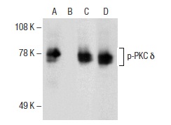
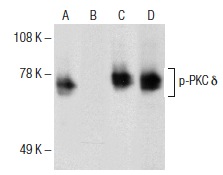
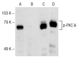
Antibodies tested include p-PKC δ (H-9): (A,B) and PKC δ (C-20): (C,D).Application
Antibodies tested include p-PKC δ (Ser 645)-R: (A,B) and PKC δ (C-20): (C,D).Application
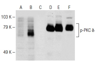
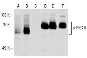
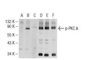
Antibodies tested include p-PKC δ (Tyr 155)-R: (A,B,C) and PKC δ (C-20): (D,E,F).Application
Antibodies tested include p-PKC δ (Tyr 565)-R: (A,B,C) and PKC δ (C-20): (D,E,F).Application
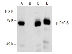
Antibodies tested include p-PKC δ (F-7): (A,B) and PKC δ (C-20): (C,D).Application
Antibodies tested include p-PKC δ (Thr 507)-R: (A,B) and PKC δ (C-20): (C,D).Application
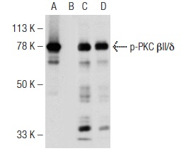
Antibodies tested include p-PKC βIl/δ (E-7): (A,B) and PKC δ (C-20): (C,D).Application
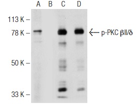
Antibodies tested include p-PKC βIl/δ (Ser 660)-R: (A,B) and PKC δ (C-20): (C,D).Application
Antibodies tested include p-PKC δ (Tyr 311)-R: (A,B,C) and PKC δ (C-20): (D,E,F).Application
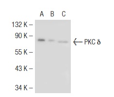
Western blot analysis of PKC δ expression in non-transfected 293T: (A), mouse PKC δ transfected 293T: (B) and NIH/3T3 (C) whole cell lysates.Application
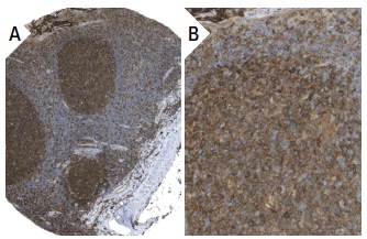
Immunoperoxidase staining of formalin fixed, paraffin-embedded human tonsil tissue showing cytoplasmic and membrane staining of lymphoid cells and squamous epithelial cells at low (A) and high (B) magnification. Kindly provided by The Swedish Human Protein Atlas (HPA) program.Application
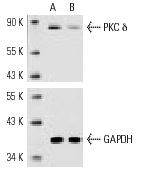
Western blot analysis of PKC δ expression in non-transfected control (A) and PKC δ siRNA transfected (B) Jurkat cells. Blot probed with PKC δ (C-20): . GAPDH (FL-335): used as specificity and loading control.Application
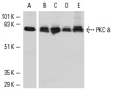
Antibodies tested include PKC δ (C-17): (A) and PKC δ (C-20): (B-E).Application
Blots were probed with p-PKC δ (Thr 507)-R: (A,B) and PKC δ (C-20)-G: (C,D).Application
Immunofluorescence staining of methanol-fixed A-431 cells showing cytoplasmic staining.Application
Western blot analysis of PKC δ expression in A-431 whole cell lysate.| Product Name | PKC δ Antibody (C-20) |
|---|---|
| Antibody Type | Primary Antibodies |
| Modification Notes | Members of the protein kinase C (PKC) family play a key regulatory role in a variety of cellular functions including cell growth and differentiation, gene expression, hormone secretion and membrane function. PKCs were originally identified as serine/threonine protein kinases whose activity was dependent on calcium and phospholipids. Diacylglycerols (DAG) and tumor promoting phorbol esters bind to and activate PKC. PKCs can be subdivided into many different isoforms (å, ∫I, ∫II, ©, ∂, é, Ω, h, †, λ/î, µ and n). Patterns of expression for each PKC isoform differ among tissues and PKC family members exhibit clear differences in their cofactor dependencies. For instance, the kinase activities of PKC ∂ and é are independent of Ca++. On the other hand, most of the other PKC members possess phorbol ester-binding activities and kinase activities. |
| Clonality | Polyclonal |
|---|---|
| Isotype | IgG |
| Host Species | Rabbit |
| Tested Applications | WBIPIFIHCELISA |
| recommended for detection of PKC &delta of mouse, rat, human and Drosophila origin by WB, IP, IF, IHC(P) and ELISA also reactive with additional species, including equine and bovine | |
| Species Reactivity | HumanMouseRat |
| Concentration | 1mg/ml |
| Purification | Affinity purified |
| Alternative Names | epitope mapping at the C-terminus of PKC &delta of human origin |
|---|---|
| Tissue Specificity | epitope mapping at the C-terminus of PKC δ of human origin |
| Entrez Gene | 5580 |
|---|
Application
Antibodies tested include p-PKC δ (H-9): (A,B) and PKC δ (C-20): (C,D).
Application
Antibodies tested include p-PKC δ (Ser 645)-R: (A,B) and PKC δ (C-20): (C,D).
Application
Antibodies tested include p-PKC δ (Tyr 155)-R: (A,B,C) and PKC δ (C-20): (D,E,F).
Application
Antibodies tested include p-PKC δ (Tyr 565)-R: (A,B,C) and PKC δ (C-20): (D,E,F).
Application
Antibodies tested include p-PKC δ (F-7): (A,B) and PKC δ (C-20): (C,D).
Application
Antibodies tested include p-PKC δ (Thr 507)-R: (A,B) and PKC δ (C-20): (C,D).
Application
Antibodies tested include p-PKC βIl/δ (E-7): (A,B) and PKC δ (C-20): (C,D).
Application
Antibodies tested include p-PKC βIl/δ (Ser 660)-R: (A,B) and PKC δ (C-20): (C,D).
Application
Antibodies tested include p-PKC δ (Tyr 311)-R: (A,B,C) and PKC δ (C-20): (D,E,F).
Application
Western blot analysis of PKC δ expression in non-transfected 293T: (A), mouse PKC δ transfected 293T: (B) and NIH/3T3 (C) whole cell lysates.
Application
Immunoperoxidase staining of formalin fixed, paraffin-embedded human tonsil tissue showing cytoplasmic and membrane staining of lymphoid cells and squamous epithelial cells at low (A) and high (B) magnification. Kindly provided by The Swedish Human Protein Atlas (HPA) program.
Application
Western blot analysis of PKC δ expression in non-transfected control (A) and PKC δ siRNA transfected (B) Jurkat cells. Blot probed with PKC δ (C-20): . GAPDH (FL-335): used as specificity and loading control.
Application
Antibodies tested include PKC δ (C-17): (A) and PKC δ (C-20): (B-E).
Application
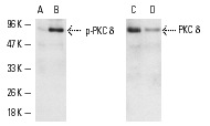
Blots were probed with p-PKC δ (Thr 507)-R: (A,B) and PKC δ (C-20)-G: (C,D).
Application
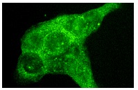
Immunofluorescence staining of methanol-fixed A-431 cells showing cytoplasmic staining.
Application
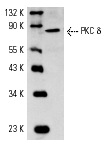
Western blot analysis of PKC δ expression in A-431 whole cell lysate.| Application Notes | recommended for detection of PKC &delta of mouse, rat, human and Drosophila origin by WB, IP, IF, IHC(P) and ELISA also reactive with additional species, including equine and bovine |
|---|
| Form | Liquid |
|---|---|
| Storage Instructions | For short-term storage, store at 4° C. For long-term storage, aliquot and store at -20ºC or below. Avoid multiple freeze-thaw cycles. |
| Storage Buffer | phosphate buffered saline , pH 7.4, 150mM NaCl, 0.02% sodium azide and 50% glycerol. |


